ЁЁ
ЁЁ
ИЮаЧзДЯИАћдкИЮдрЗЂг§ЁЂдйЩњКЭИЮАЉаЮГЩжаЕФзїгУ
Hepatic stellate cells in liver development, regeneration, and cancer
ЁЁ
Chunyue Yin,1 Kimberley J. Evason,2 Kinji Asahina,3 and Didier Y.R. Stainier1
First published May 1, 2013 - More info
ЁЁ
Abstract
Hepatic stellate cells are liver-specific mesenchymal cells that play vital roles in liver physiology and fibrogenesis. They are located in the space of Disse and maintain close interactions with sinusoidal endothelial cells and hepatic epithelial cells. It is becoming increasingly clear that hepatic stellate cells have a profound impact on the differentiation, proliferation, and morphogenesis of other hepatic cell types during liver development and regeneration. In this Review, we summarize and evaluate the recent advances in our understanding of the formation and characteristics of hepatic stellate cells, as well as their function in liver development, regeneration, and cancer. We also discuss how improved knowledge of these processes offers new perspectives for the treatment of patients with liver diseases.
ЁЁ
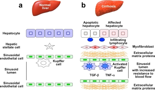
Hepatic stellate cells are located in the space of Disse between the sinusoidal endothelial cells and hepatic epithelial cells, and account for 5%ЈC8% of the cells in the liver. In a healthy liver, stellate cells are quiescent and contain numerous vitamin A lipid droplets, constituting the largest reservoir of vitamin A in the body (reviewed in ref. 1). When the liver is injured due to viral infection or hepatic toxins, hepatic stellate cells receive signals secreted by damaged hepatocytes and immune cells, causing them to transdifferentiate into activated myofibroblast-like cells (reviewed in ref. 2). As the primary extracellular matrixЈCproducing (ECM-producing) cells in liver, activated stellate cells generate a temporary scar at the site of injury to protect the liver from further damage. In addition, hepatic stellate cells secrete cytokines and growth factors that promote the regeneration of hepatic epithelial cells. In chronic liver disease, prolonged and repeated activation of stellate cells causes liver fibrosis, as characterized by widespread scar formation and perturbation of liver architecture and function (reviewed in ref. 3). Recent clinical and experimental evidence indicates that hepatic fibrosis is reversible upon removal of the underlying etiological agent (4ЈC6). During the regression of liver fibrosis, the number of activated hepatic stellate cells is greatly reduced by the induction of cellular senescence and apoptosis, or by the return to the quiescent state (2, 5ЈC7). Because of their pivotal roles in liver repair and disease pathogenesis, hepatic stellate cells have been a major focus of liver research. However, our knowledge of their cell biology is far from complete, mainly due to the challenges of studying these cells in vivo.
ЁЁ
This Review focuses on the recent insights and emerging investigations into the formation of hepatic stellate cells and their function in liver development, regeneration, and hepatocellular carcinoma (HCC). The regulation of stellate cells in liver fibrosis as well as the design of antifibrotic therapies is reviewed separately in this issue (8).
JCI - Hepatic stellate cells in liver development, regeneration, and cancer https://www.jci.org/articles/view/66369
ЁЁ
ЁЁ
.png)
.png)
