匹兹堡大学 欧米茄-3脂肪酸抑制肝癌细胞的生长
University of Pittsburgh:Omega-3 Fatty Acids Inhibit Growth of Liver Cancer Cells
匹兹堡大学(University of Pittsburgh)一个研究小组的两项新研究表明,omega-3脂肪酸——一种存在于鱼油、某些种子和坚果中的高浓度物质——可以显著抑制肝癌细胞的生长。这些研究是在华盛顿举行的美国癌症研究协会年会上发表的,提示omega-3脂肪酸可能是治疗和预防人类肝癌的有效疗法。
肝细胞癌占所有肝癌的80%到90%,通常在确诊后三到六个月内死亡。
第一项研究考察了欧米茄-3和欧米茄-6多不饱和脂肪酸在人肝癌细胞中的作用和作用机制。肝细胞癌占所有肝癌的80%到90%,通常在确诊后三到六个月内死亡。
人们早就知道欧米茄-3脂肪酸可以抑制某些癌细胞。因此,我们感兴趣的是确定这些物质是否能抑制肝癌细胞。如果是这样,我们还想知道这种抑制发生的机理,Wu Tong博士是匹兹堡大学医学院(University of Pittsburgh School of Medicine)移植病理学部的一名成员。
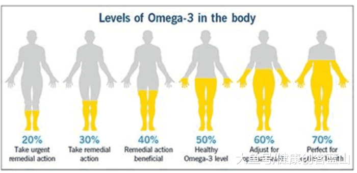
OMEGA-3 ACID IN THE BODY
研究人员用欧米伽-3脂肪酸二十二碳六烯酸(DHA)和二十碳五烯酸(EPA)或欧米伽-6脂肪酸花生四烯酸(AA)治疗肝细胞癌细胞12至48小时。DHA和EPA处理对细胞生长有剂量依赖性抑制作用,而AA处理无显著影响。
据研究人员称,omega-3脂肪酸对癌细胞的影响可能是由于诱导细胞凋亡或程序性细胞死亡。实际上,研究人员发现,DHA的治疗可以诱导细胞核内一种酶的分裂或分裂,这种酶被称为聚(ADP -核糖)聚合酶(PARP),参与修复DNA损伤、介导细胞凋亡和调节免疫反应。这种酶的裂解被认为是细胞凋亡的一个指标。此外,DHA和EPA的治疗间接降低了另一种被称为-beta-catenin的蛋白水平,这种蛋白的过量与各种肿瘤的发生有关。
“已知beta-catenin可以促进细胞生长,也与肿瘤细胞的促进有关。因此,我们发现欧米茄-3脂肪酸可以降低-beta-catenin的水平,这进一步证明了这些化合物有能力与参与肿瘤进展的几个途径相互作用。
在第二项研究中,研究人员用omega-3和omega-6脂肪酸治疗胆管癌肿瘤细胞12到48小时。胆管癌是一种恶性程度特别高的肝癌,发生在从肝脏携带胆汁的导管中,死亡率极高。
同样,欧米伽-3脂肪酸DHA和EPA的治疗对癌细胞生长有剂量依赖性抑制作用,而欧米伽-6脂肪酸AA治疗没有显著效果。同样,DHA治疗可诱导胆管癌细胞PARP分裂,DHA和EPA治疗可显著降低细胞内beta-catenin水平。
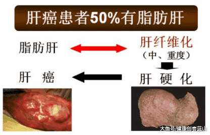
Wu博士说,这些发现表明, 欧米茄- 3脂肪酸不仅可能是一种有效的人类肝癌的治疗,但也可以是预防脂肪肝的一种手段, 脂肪肝是一种慢性疾病,特征是脂肪在肝脏积聚,并被认为是肝细胞癌的前兆。他说,这个过程的下一步是测试欧米茄-3脂肪酸在含有人类肝脏肿瘤的老鼠体内的效果。
来源:
摘要2679,2680,美国癌症研究协会(AACR)在华盛顿会议中心举行的年度会议2006年4月,
关键概念:-3脂肪酸,EPA, DHA,肝癌,肝细胞癌,胆管癌
欧米茄-3脂肪酸可以抑制肝癌细胞的生长
Two new studies by a University of Pittsburgh research team suggest that omega-3 fatty acids--substances that are found in high concentrations in fish oils and certain seeds and nuts--significantly inhibit the growth of liver cancer cells. The studies, presented at the annual meeting of the American Association for Cancer Research, in Washington, D.C., suggest that omega-3 fatty acids may be an effective therapy for both the treatment and prevention of human liver cancers.
The first study, looked at the effect and mechanism of omega-3 and omega-6 polyunsaturated fatty acids in human hepatocellular carcinoma cells. Hepatocellular carcinoma accounts for 80 to 90 percent of all liver cancers and is usually fatal within three to six months of diagnosis.
"It has been known for some time that omega-3 fatty acids can inhibit certain cancer cells. So, we were interested in determining whether these substances could inhibit liver cancer cells. If so, we also wanted to know by what mechanism this inhibition occurs," said Tong Wu, M.D., Ph.D., a member of the division of transplantation pathology, University of Pittsburgh School of Medicine, in whose laboratory the research was conducted.
The investigators treated the hepatocellular carcinoma cells with either the omega-3 fatty acids docosahexaenoic acid (DHA) and eicosapentaenoic acid (EPA) or the omega-6 fatty acid arachidonic acid (AA), for 12 to 48 hours. DHA and EPA treatment resulted in a dose-dependent inhibition of cell growth, whereas AA treatment exhibited no significant effect.
According to the investigators, the effect of omega-3 fatty acids on cancer cells likely is due to the induction of apoptosis, or programmed cell death. Indeed, the investigators found that DHA treatment induced the splitting up, or cleavage, of an enzyme in the cell nucleus known as poly (ADP-Ribose) polymerase, or PARP, which is involved in repairing DNA damage, mediating apoptosis and regulating immune response. The cleavage of this enzyme is considered a tell-tale indicator of apoptosis. Furthermore, DHA and EPA treatment indirectly decreased the levels of another protein known as beta-catenin, an overabundance of which has been linked to the development of various tumors.
"Beta-catenin is known to promote cell growth and also is implicated in tumor cell promotion. Therefore, our finding that omega-3 fatty acids can decrease levels of beta-catenin is further evidence that these compounds have the ability to interact on several points of pathways involved in tumor progression," explained Dr. Wu.
In the second study, Abstract number 2680, the investigators treated cholangiocarcinoma tumor cells with omega-3 and omega-6 fatty acids for 12 to 48 hours. Cholangiocarcinoma is a particularly aggressive form of liver cancer that arises in the ducts that carry bile from the liver and has an extremely high mortality rate. Again, the omega-3 fatty acids DHA and EPA treatments resulted in a dose-dependent inhibition of cancer cell growth, while the omega-6 fatty acid AA treatment had no significant effect. Likewise, DHA treatment induced a cleavage form of PARP in cholangiocarcinoma cells, and DHA and EPA treatment significantly decreased the level of beta-catenin protein in the cells.
According to Dr. Wu, these findings suggest that omega-3 fatty acids not only may be an effective therapy for the treatment of human liver cancers but may also be a means of protecting the liver from steatohepatitis, a chronic liver disease characterized by the buildup of fat in the liver and believed to be a precursor of hepatocellular carcinoma. The next step in the process, he said, is to test the effects of omega-3 fatty acids in mice harboring human liver tumors.
Source:
Abstract number 2679, 2680, Annual Meeting of the American Association for Cancer Research (AACR), at the Washington Convention Center in Washington, D.C., April 2006
Key concepts: omega-3 fatty acids, EPA, DHA, liver cancer, hepatocellular carcinoma, cholangiocarcinoma
Omega-3 Fatty Acids Inhibit Growth of Liver Cancer Cells
http://www.rejuvenation-science.com/research-news/omega-3-fish-oil-1/n-omega-3-liver-cancer


.png)