��
Psoriasis Studies~Inside out disease
Features of psoriasis~ proliferation,inflammation, scales,itching
cell types in psoriasis lesion~Th17 predominant, Th1
��ͻ״ϸ�� Dendritic Cells ~cellular metabolism shapes the functional properties of DCs.
Dendritic cells,Macrophages, Cytokine-signaling in Psoriasis
Mast cells~ are main source of IL-17 in psoriasis lesions. mast cells, but not T cells or macrophages, were the predominant cell type producing IL-17 in psoriasis lesions.
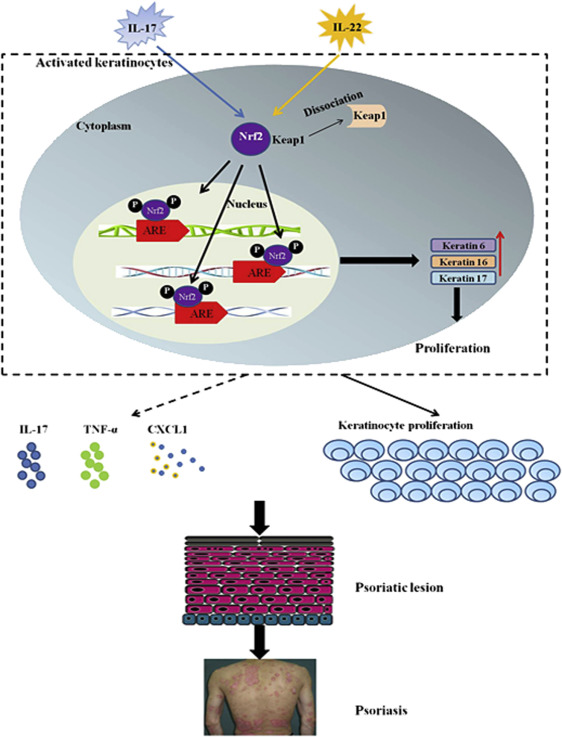
Nrf2 was overexpressed and activated in the epidermis of psoriatic lesions. K6, K16, K17 Hallmarks of psoriasis
Proliferation pathway~ DC, IL17, IL22 -Nrf2-K6, K16, K17Sequences of immune response
roles of phosphodiesterase type 4 (PDE-4) and its inhibitor
Bruton's tyrosine kinase inhibitor suppresses imiquimod-induced psoriasis-like inflammation. Bruton's tyrosine kinase (BTK) has been reported to execute important signaling functions in innate immune cells such as dendritic cells (DCs) and gamma delta T cells.
Role of TGF-betta in kiratinocyte profliferaiton
activated immune cells undergo anaerobic glycosis
metabolism and proinflammatory/antiinflammatory cytokine production
activation of dendritic cells via TLRs~Activation of DCs via TLRs promotes significant upregulation of aerobic glycolysis, which regulates the immune function of both human and mouse DCs; Dendritic cells are what they eat: how their metabolism shapes T helper cell polarization
PI3K/ART/mTOR pathways
herbal ingredients for treating psoriasis~ danshensu, luoteolin,
Natural treatments: UVR
Autoantigens~
LPS/TLRs signaling pathway~TLR4
Effect of fatty acids on TLRs signaling pathway
Roles of IgE, IgE receptors on mast cells and B cells
sources of IL-23~ mature dendritic cells
sources of IL-17~ Th17, CD8+ effector T cells
PRO-inflammotory cytokines: IL-1,IL-6��IL-23/17, TNF-alpha
IL-17A stimulated keratinocytes activated PI3K/AKT/mTOR signaling and inhibited autophagy by simultaneously inhibiting autophagosome formation and enhancing autophagic flux.
Vitamin c~glycolysis,inflammation~ NF-kB,Nrf2,TNFa, EGFR? natural antihistamine
Inhibition of glycolysis (GAPDH) that mature dentrictic/M1 macrophages/effector T cells rely on by vitamin C
Vitamin C blocks TNF-alpha-induced NF-kappaB activation
Vitamin C blocks TNF-alpha-induced NF-kappaB activation and ICAM-1 expression in human neuroblastoma cells
Vitamin C blocks TNF��-induced NF k B activation and ICAM-1 expression in human neuroblastoma cells
Vitamin c~activation of AMPK, while AMPK inhibits mTOR
vitamin C inhibited the TNF-��-induced activation of not only the mitogen-activated protein kinase (MAPKs), but also nuclear factor-kappa B (NF-��B) signaling.
Inhibition/down-regulate PI3K/ART-mTOR signaling BY EPA, DHA (PTEN), EGCG,
AA is a competitive inhibitor of adenylate cyclase~anti-proliferation?
Natural kinase inhibitors:
Polyphenol Analogues~phenolic acids (tannic, gallic, and ellagic acid.), flavonoids, anthraquinones, coumarins, and lignans. EGCG- inhibits basophils to release histamine
gallic acid~ reduced IL-6 gene expression,
Glycyrrhizic acid~ as HMGB1 inhibitor (TLR2, TLR4, RAGE), mast cell stabilizer, reducing B cell to produce IgE, suppress IL-4, restore immune balance of Th1/Th2.
GA inhibits TLR2 activation
HMGB1: The Central Cytokine for All Lymphoid Cells
Glycyrrhizin ameliorates experimental colitis through attenuating interleukin-17-producing T cell responses via regulating antigen-presenting cells
Glycyrrhizin enhances interleukin-10 production by liver dendritic cells in mice with hepatitis
Glycyrrhizin GL inhibited the activation of the NF‑��B and MAPK/ERK signaling pathways induced by HMGB1 and decreased the expression of monocyte chemoattractant protein‑1 (MCP‑1) and myeloid cell leukemia 1 (Mcl‑1).Flavonoid Analogues
Berberine/huangliansu/danshensu~Berberine/danshensu inhibits the expression of TNFalpha, MCP-1, and IL-6 in AcLDL-stimulated macrophages through PPARgamma pathway
Lactic acid~ inhibits glycolysis (chelator of nickle)~LA significantly suppressed LPS-induced cytokine production and NF-��B transcriptional activity in mouse bone marrow-derived mast cells and cytokine production in peritoneal mast cells. Mostly, lactate is recognized as a molecule capable of suppressing immune responses, through inhibition of T cells, Mϕs, and dendritic cells.
Lactic Acid Reduces LPS-Induced TNF-�� and IL-6 mRNA Levels Through Decreasing IKB�� Phosphorylation
Lactate Suppresses Macrophage Pro-Inflammatory Response to LPS Stimulation by Inhibition of YAP and NF-��B Activation via GPR81-Mediated Signaling
Magnessium~ hypomagnesemia could induce a release of histamine from dermal mast cells. A characteristic allergy-like crisis occurs spontaneously in Mg-deficient rats, the first visible symptom being a peripheral vasodilatation of the ears. A low Mg medium (0.2 mM Mg) induced much more histamine release from mast cells. Magnesium is a natural calcium antagonist. Calcium is essential for Th17 activation. We localized Th17 cells predominantly to the dermis of psoriasis skin lesions, confirmed that IL-17 mRNA increased with disease activity, and demonstrated that IL-17 mRNA expression normalized with cyclosporine (a calcium dependant calcineurin inhibitor) therapy.
Lauric acid ameliorates lipopolysaccharide (LPS)-induced liver inflammation by mediating the TLR4/MyD88 pathway in Sprague Dawley (SD) rats. CONTROVERSY studies also found lauric acid stimulate TLR4
Iowa university~GML inhibits TLR2 signaling
Induction of proinflammatory cytokines by long-chain saturated fatty acids in human macrophages
Palmitate and insulin synergistically induce IL-6 expression in human monocytes
linseed oil/vitamin c for psoriasis~ VC inhibits GAPDH, oxVC/DHA inhibits NFkB, mTOR
Omega-3 polyunsaturated fatty acid promotes the inhibition of glycolytic enzymes and mTOR signaling by regulating the tumor suppressor LKB1
HIF1�� ��-VEGF) expression is required for the differentiation of Th17 cells
Omega-3 polyunsaturated fatty acid promotes the inhibition of glycolytic enzymes and mTOR signaling by regulating the tumor suppressor LKB1
ALA (alpha-linolenic acid) intake not only significantly reduced tumor necrosis factor-�� (TNF-��) and interleukin-6 (IL-6) concentrations. ALA significantly inhibited the secretion of proinflammatory cytokines including tumor necrosis factor-�� (TNF-��), interleukin-6 (IL-6) and interleukin-1�� (IL-1��) and increased anti-inflammatory cytokine. ALA significantly inhibited the phosphorylation of I��B�� and NF-��B (p65) activation in ALI. ALA showed anti-inflammatory effects in mice with LPS-induced ALI. treatment with a high concentration of ALA markedly increased the production of IL-10. ALA suppressed the activation of NF-��B in LPS-induced ALI.
ALA on mitochondrial respiration~FAO (1) The overall beta-oxidation in total mitochondria was in the order C18:3, n-3 greater than C18:2, n-6 greater than C18:1, n-9, independent of the amount of albumin in the medium.
LKB1 restrains dendritic cell function~LKB1 orchestrates dendritic cell metabolic quiescence and anti-tumor immunity. LKB1 is potentiated by AMPK (which is potentiated by vitamin c)
the capacity of DHA to activate the AMPK signalling and negatively regulate the HIF-1�� functions.
Roles of HIFa~ HIF1��, a transcription factor that controls the cellular response to hypoxia, activates the glycolytic pathway and, as such, promotes inflammation 47. HIF1�� expression is required for the differentiation of Th17 cells 45, a T cell subset expanded in many autoimmune and inflammatory diseases. HIF1���\inhibitor echinomycin reduced Th1 and Th17 responses, and attenuated a mouse model of acute graft�\versus�\host disease
Natural antihistamine~vitamin c, glycyrrhzin, quercetin, EGCG, magnesium?
EGCG inhibits mTOR signaling pathway~ Epigallocatechin-3-gallate (EGCG) is an ATP-competitive inhibitor of PI3K and mTOR with Ki values around 300 nM. EGCG inhibits cell proliferation and AKT phosphorylation at Ser473 in MDA-MB-231and A549 cells. Molecular docking studies show that EGCG binds well to the PI3K kinase domain active site.
EGCG could significantly impede expressions of HIF-1�� and VEGF proteins.EGCG affects human dendritic cell differentiation and maturation~induced apoptosis in DC and Most importantly, mature DCs treated with EGCG inhibited stimulatory activity toward allogeneic T cells while secreting high amounts of IL-10. EGCG induces immunosuppressive alterations on human MODCs, both by induction of apoptosis and suppression of cell surface molecules and antigen presentation.
Epigallocatechin-3-gallate (EGCG) inhibits imiquimod-induced psoriasis-like inflammation of BALB/c mice
Epigallocatechin gallate induces apoptosis of monocytes
EGCG induced a reduction in proliferation of autoreactive T cells, production of proinflammatory cytokines, and Th1 and Th17 subpopulations, and an increase in regulatory T-cell populations.
EGCG inhibits proliferation of gastric cancer, pancreatic cancer cells
RETINOIC ACID
retinoic acid inhibiting the differentiation of dendritic cells, maturation and induction of the T-helper cell type-2 response. All-trans-retinoic acid atRA can inhibit the maturation of dendritic cells derived from cord blood mononuclear cells and that the effect can be stopped by a selective RAR�� antagonist, Ro 41-5253.atRA reduced the ability of dendritic cells to induce allogeneic T cells in mixed lymphocyte reaction. Similarly, Ro 41-5253 can reverse this reduction. atRA can affect dendritic cell-directed Th immune reaction and can promote Th2 response. The action may be mediated by RAR
Retinoic acid promotes the development of Arg1-expressing dendritic cells for the regulation of T-cell differentiation
Hypericin~a natural photosensitizer/vitamin c, omega-3, iron ~inhibition of HIF-1.
Conclusion: The present study demonstrated a marked increase of HIF- 1 alpha immunoreactivity in psoriatic skin.
Hypoxia-inducible factor-1�� (HIF-1��) is a more important factor in psoriatic epidermal proliferation. Study on HIF-1�� Gene Translation in Psoriatic Epidermis with the Topical Treatment of Capsaicin Ointment
https://www.ncbi.nlm.nih.gov/pmc/articles/PMC3263732/��
Pathomechanism�Ľǻ���
��Ƥ�ɻ��ײ㡢���㡢������ͽ��ʲ���ɡ�����м��Ƥ���У������γ�ϸ���ӻ��ײ㵽�����ת��ʱ����������Ƥ���Լ13��������48Сʱ��Ҳ�б����ƣ�����м��Ƥ���У�ϸ�����ڴ�����Ƥ���е�311Сʱ���̵����������γ�ϸ����36Сʱ���������м��Ƥ���н����γ�ϸ����ֳ���Լӿ졣��м���IJ������Ʊ���Ϊ��ϸ����ֳ���ٺͽ����γ�ϸ���ӻ��ײ�����������Ǩ���йء������γ�ϸ������ֳ�ܵ����ַ��ӵĵ��أ��绷״������(AMP)������øC����֬øC��ת���������ө\����
Figure 1. Role of mTOR and cellular metabolism in the regulation of MHC II-dependent antigen presentation by DCs.
Metabolic pathways and upstream signaling pathways regulating these are indicated in red and black, respectively. The left and the right sides of the figure depict how MHC II-dependent antigen presentation is metabolically regulated in unactivated (immature) and TLR-stimulated (mature) DCs, respectively.Dendritic cells are what they eat: how their metabolism shapes T helper cell polarization - ScienceDirect
https://www.sciencedirect.com/science/article/pii/S0952791518301080
��Previous studies showed that active oxidative phosphorylation in mitochondria is associated with immature or tolerogenic DCs, while increased glycolysis upon pathogen sensing can promote immunogenic DC functions.
Metabolic Control of Dendritic Cell Functions: Digesting Information - PubMed
https://pubmed.ncbi.nlm.nih.gov/31073300/��
��
��
Psoriasis pathogenesis and the development of novel targeted immune therapies - PubMed
https://pubmed.ncbi.nlm.nih.gov/28887948/#&gid=article-figures&pid=figure-1-uid-0��
https://onlinelibrary.wiley.com/doi/full/10.1111/1346-8138.14139
��
Main axis of the pathogenesis of psoriasis. Cells important in the pathogenesis of psoriasis are dendritic cells (DC), T�\helper (Th)17 (and Th1), and keratinocytes. DC activated by various stimuli excessively produce and secrete tumor necrosis factor (TNF)�\�� and interleukin (IL)�\23 (including IL�\12). IL�\23 induces differentiation of naive T cells into Th17. Activated Th17 cells overproduce IL�\17 and IL�\22. TNF�\�� and IL�\17 activate keratinocytes, promote epidermal hyperplasia, recruit inflammatory cells, such as neutrophils, and induce antimicrobial peptide (AMP) production. IL�\12 produced by dendritic cells also induces Th1, and Th1 produce cytokines, including interferon (IFN)�\��. This irregular immune response continues as TNF�\�� activates dendritic cells. CXCL, chemokine (C�\X�\C motif) ligand.
The close relation between IL�\17 and psoriasis was reported. High levels of IL�\17 mRNA were detected in psoriatic lesional skin, but not in non�\lesional skin.25 In keratinocytes, IL�\17 enhanced the expression of IL�\6 and IL�\8, which are known as pro�\inflammatory cytokines and exacerbate psoriasis.26 In addition, topical application of imiquimod, a Toll�\like receptor (TLR)7/8 ligand and potent immune activator, induced psoriasis�\like dermatitis in mice together with the expression of IL�\17A and IL�\17F.27 Furthermore, both CsA and anti�\TNF�\�� agents decreased the levels of IL�\17A, IFN�\��, IL�\23p19 and chemokine (C�\C motif) ligand 20 in psoriatic lesions in conjunction with improvement of psoriatic eruptions, indicating that pro�\inflammatory cytokines, including IL�\17A, are involved in the development of psoriasis.26, 28-30 These findings suggest that the IL�\17 family plays an important role in psoriasis.
Pathogenesis of psoriasis and development of treatment - Ogawa - 2018 - The Journal of Dermatology - Wiley Online Library
https://onlinelibrary.wiley.com/doi/full/10.1111/1346-8138.14139��
Enhanced glucose uptake and a switch to glycolysis are key traits of M1 macrophages, whereas enhanced fatty acid oxidation and oxidative phosphorylation are the main metabolic characteristics of M2 macrophages.
Glycolytic Stimulation Is Not a Requirement for M2 Macrophage Differentiation - ScienceDirect
https://www.sciencedirect.com/science/article/pii/S1550413118305138��
https://www.frontiersin.org/articles/10.3389/fimmu.2014.00491/full
��
��
https://openi.nlm.nih.gov/detailedresult.php?img=PMC2582809_ar2413-1&req=4
��
http://link.springer.com/chapter/10.1007/978-3-319-19530-8_9
��
https://www.mdpi.com/1420-3049/24/11/2157
��
��
http://miriam-english.org/files/Harnessing_Infection_to_Fight_Cancer.html
��
��
http://journal.frontiersin.org/article/10.3389/fimmu.2013.00082/full
��
https://www.frontiersin.org/articles/10.3389/fimmu.2018.03176/full
http://www.pnas.org/content/99/1/351
��
��
https://computing.dcu.ie/~hruskin/hruskin.html
��
The differentiation of Th17 cells is supported by IL-6 and TGF-��, whereas IL-23, IL-1��, and IL-21 promote their expansion (2�C4).
Interestingly, polymorphisms at loci encoding components of IL-23 and its receptor��IL23A, IL12B, and IL23R��have been associated with an increased risk of developing psoriasis (5�C9). Psoriasis lesions contain increased amounts of IL-17 mRNA and increased numbers of Th17 cells (2, 10, 11). Similarly, increased tissue expression of IL-17 and numbers of Th17 cells are seen in rheumatoid arthritis, Crohn��s disease, autoimmune uveitis, lupus erythematosus, ankylosing spondylitis, asthma, and multiple sclerosis (12). The pathophysiologic relevance of the IL-23�CIL-17 axis in autoinflammatory disease is highlighted by the clinical effectiveness of Abs targeting IL-23/IL-12 p40 and IL-17 in treating psoriasis as well as the other aforementioned diseases (13�C16).Surprisingly, we observe that most IL-17+ cells in normal and psoriatic skin are mast cells, not T cells. Interestingly, mast cell numbers are decreased in psoriasis lesions after successful treatment with anthralin, psoralen plus UVA light therapy, or cyclosporine (25�C27). Neutrophils also are enriched in psoriasis lesions, especially in the epidermis where they aggregate in Munro��s microabscesses (MMs) in the stratum corneum and spongiform pustules of Kogoj (SPKs) in the stratum spinosum (28).
IL-17 orchestrates innate immune responses against extracellular pathogens by inducing expression of antimicrobial peptides (AMPs) and neutrophil-tropic chemokines CXCL1, CXCL2, and IL-8 (32�C35). The same AMPs and chemokines are found at extremely high levels in psoriatic epidermis (36�C38). Not surprisingly, mice and humans with deficits in IL-17 production or signaling are highly susceptible to infection with extracellular bacteria and fungi (39�C42).
effective antimicrobial activity by neutrophils and mast cells depends on the formation of structures called extracellular traps (ETs), termed NETs and MCETs, respectively (43�C45). ETs are formed through a specialized process of cell death termed ETosis (46), where chromatin extends into fine, weblike threads to which proteins are bound. In particular, NETs can contain myeloperoxidase (MPO), proteinase 3, and AMPs such as cathelicidin (LL-37) (47). The process of NET formation (NETosis) can be triggered by extracellular bacteria and fungi or their components (48, 49). MCETs contain tryptase, LL-37, and chromatin (45), forming in response to bacteria, H2O2, or PMA (45, 48). In humans, NETs have been visualized in physiologic host responses to infections (44, 50) and have been implicated in the pathology of anti-neutrophil cytoplasmic Ab-induced vasculitis (51). Additionally, a recent study showed that lupus nephritis is associated with an inability to degrade NETs in blood (52).
We observe that mast cells and neutrophils release IL-17 into the skin though ETosis as well as conventional degranulation.
http://www.jimmunol.org/content/187/1/490
��
A positive feedback loop. Mast cell activation by cytokinergic IgE induces cytokine secretion by mast cells in the absence of antigen. The cytokines stimulate mast cell survival and class switching to IgE in B cells. Continued production of IgE and cytokines occurs in the absence of antigen.
Some 10 years ago it emerged that at sufficiently high concentrations certain monoclonal mouse IgEs exert previously unsuspected effects on mast cells. Thus they can both promote survival and induce activation of mast cells without the requirement for antigens. This was a wake up call that appears to have been missed (or dismissed) by the majority...
... DNCB activates immunity to release IgE from the plasma B cells, similar to the immune defense against parasites but it is associated with over-activating immunity. IgE production results in the degranulation of activating mast cells to release histamine which causes itching and edema of the skin [38]. STB, AOM and the mixture of STB and AOM alleviated the clinical AD symptoms to similar levels as the Normal-Con mice by reducing serum IgE concentrations and mast cells in the skin tissues. ...
. complex of RAG1 and RAG2 can itself initiate the process of receptor revision, since all the other machinery for repairing DNA breaks in cells is constitutive (Nemazee, 2006). RAG genes are normally silenced once a func- tional Ig is expressed in B cells, but may be re-expressed in the presence of IL-6 (Hillion et al., 2007a,b). IL-6 secretion is com- monly observed when mast cells are stimulated by cytokinergic IgE; when this occurs in the target organs of allergy, it may lead to the local expression of polyspecific/cytokinergic IgE. ...
View in full-text
How IgE mediates an allergic reaction via interaction with its two receptors. (Left) Interactions of membrane-bound IgE (mIgE, blue) with CD23 (tangerine) on B-cells regulates soluble IgE (sIgE) production. (Right) Cross-linking of IgE bound to Fc��RI (scarlet) on mast cells or basophils by allergens (brown) triggers the release of mediators, causing allergy.
IgE is the antibody isotype found at the lowest concentration in the circulation. However IgE can undeniably play an important role in mediating allergic reactions; best exemplified by the clinical benefits of anti-IgE monoclonal antibody (omalizumab) therapy for some allergic diseases.
��
Mast cell sensitization is initiated by exposure to an allergen. In this case, a pollen grain allergen crosses the epithelial barrier and activates the production of IgE antibodies from B cells. The IgE bind surface high affinity receptors (FcRI) on the surface of mast cells and arm them for subsequent exposure.
... Crosslinking of IgE bound to mast cells by allergens, in turn, triggers the release of mediators, such as histamine and leukotrienes that are responsible for arteriolar dilation, increased vascular permeability, itching, rhinorrhea, mucus secretion, and smooth muscle contraction in the lung. The mediators and cytokines released during the early phase of an immune response to an inciting allergen trigger a further cellular inflammatory response over the next 4-8 h (late-phase inflammatory response) resulting in recurrent symptoms (usually nasal congestion) that often persist [40][41][42]. A lot of medical treatment modalities used as a treatment of AR, such as antihistamines, steroids, montelukast (Singulair), and immunotherapy.
Quercetin is a naturally occurring polyphenol flavonoid which is rich in antioxidants. It has anti-allergic functions that are known for inhibiting histamine production and pro-inflammatory mediators. Quercetin can regulate the Th1/ Th2 stability, and decrease the antigen-specific IgE antibody releasing by B cells. Quercetin has a main role in anti-inflammatory and immunomodulatory function which makes it proper for the management of different diseases. Allergic diseases are a big concern and have high health care costs. In addition, the use of current therapies such as ß2-agonists and corticosteroids has been limited for long term use due to their numerous side effects. Since the effect of quercetin on allergic diseases has been widely studied, in the current article, we review the effect of quercetin on allergic diseases, such as allergic asthma, allergic rhinitis (AR), and atopic dermatitis (AD).
��
Upon activation, naïve CD4+ T cells differentiate into a number of specialized T helper (Th) cell subsets. Th2 cells are central players in immunity to helminths and are implicated in mediating the inflammatory pathology associated with allergies. https://pubmed.ncbi.nlm.nih.gov/31611881/
��
Figure 1. Allergy response pathway. As part of an allergic response, allergens are presented to naïve T cells, which activates Th2 cells in the presence of IL-4. Th2 cells then release cytokines including IL-3, IL-4 and IL-13. This in turn activates the release of IgE-producing antibodies as well as activating eosinophils and mast cells. Figure adapted from Fujita H, Meyer N, Akdis M et al (2012).
Memory Th2 cell responses are recognised as a significant contributor of exacerbated allergic diseases. Th2 cells comprise a core part of the adaptive immune system. It has been proposed that minimal bacterial exposure early in life shifts the balance of the Th1/Th2 ratio towards a dominant Th2 immune response. Th2 cells secrete cytokines including IL-4, IL-5, IL-6 and IL-13. This in turn activates IgE antibody production, as well as mast cells and eosinophils (Figure 1).
https://www.esgct.eu/Blog/Treating-allergy-with-a-single-vaccine.aspx
Th2 Cell - an overview | ScienceDirect Topics
https://www.sciencedirect.com/topics/medicine-and-dentistry/th2-cell
Cytokines such as IL-4 and IL-5 released by Th2 cells stimulate, respectively, B-cell switching to the production of IgE antibody and activation of eosinophils. The coordinate actions of these effector mechanisms result in heightened immunity against, for example, helminthic parasites, which can be coated with IgE and destroyed by the toxic granular contents of eosinophils.��
LKB1 signaling in dendritic cells limits their T cell-activating potential. LKB1 is phosphorylated in DCs in the tumor microenvironment, while it is depleted by LPS or E. coli favoring Treg expansion. LKB1 limits the ability of DCs to induce T cell priming by repressing a variety of activating pathways. These effects lead to LKB1-deficient DCs to promote dysregulated T cell effector activity, with predominant increase in thymus-derived regulatory T cell priming but also increased priming of pro-immunogenic effector Th17, Th1 and CD8+ T cells. Mechanistically, upon loss of LKB1, DCs enhance their expression of MHC molecules, co-stimulatory molecules (e.g., CD86, OX40L), cytokines (e.g., IL-6, IL-2) and migration receptors (e.g., CCR7) �� all of which contribute to enhanced T cell priming. The predominant activation of regulatory T cells and Th17 cells upon LKB1 deletion in DCs contributes to tumor growth
Overall, LKB1 emerges as a fundamental regulator of the core DC function to control T cell responses and maintain their immunological quiescence, at least partially via limiting DC migration, co-stimulatory molecule (CD86 and OX40L) and cytokine (IL-6) expression (Fig. 1).9�C11 LKB1 loss in DCs results in their uncontrolled stimulation of T cells, foremost of Tregs by cDC2s in the thymus and periphery as well as peripheral Th17 cells. Prevention of mTOR signaling in DCs, likely in concert with limiting glycolytic metabolism,2�C4 appears to account for aspects of LKB1-mediated regulation of T cell immunity by DCs, such as Treg homeostasis.
LKB1 restrains dendritic cell function
https://www.ncbi.nlm.nih.gov/pmc/articles/PMC6796839/��
http://www.jimmunol.org/content/198/6/2223
��
Nrf2 Promotes Keratinocyte Proliferation in Psoriasis through Up-Regulation of Keratin 6, Keratin 16, and Keratin 17 - Journal of Investigative Dermatology
https://www.jidonline.org/article/S0022-202X(17)31561-0/fulltext��
��
Jounard of Investigation Dermatology. 2008 May;
Psoriasis vulgaris lesions contain discrete populations of Th1 and Th17 T cells
1Laboratory for Investigative Dermatology, The Rockefeller University, New York, New York 10021, USA.
The importance of T helper 17 (Th17) cells in inflammation and autoimmunity is now being appreciated. We analyzed psoriasis skin lesions and peripheral blood for the presence of IL-17-producing T cells. We localized Th17 cells predominantly to the dermis of psoriasis skin lesions, confirmed that IL-17 mRNA increased with disease activity, and demonstrated that IL-17 mRNA expression normalized with cyclosporine therapy. IL-22 mRNA expression mirrored IL-17 and both were downregulated in parallel with keratin 16. Th17 cells are a discrete population, separate from Th1 cells (which are also in psoriasis lesions), and Th2 cells. Our findings suggest that psoriasis is a mixed Th1 and Th17 inflammatory environment. Th17 cells may be proximal regulators of psoriatic skin inflammation, and warrant further attention as therapeutic targets.Ѱ������м�����������ɢ��Th1��Th17 Tϸ��Ⱥ
T helper 17 (Th17)ϸ������֢�����������е���Ҫ�����ڵõ����ӡ����Ƿ�������м��Ƥ�����˺�����Ѫ���Ƿ����il -17����Tϸ�������ǽ�Th17ϸ����Ҫ��λ����м��Ƥ�����Ƥ��֤ʵIL-17 mRNA�漲��������ӣ���֤ʵ�ڻ�����(cyclosporine)���ƺ�IL-17 mRNA������������IL-22 mRNA������IL-17һ�£�����ǵ���16ƽ���µ���Th17ϸ����һ����ɢ��Ⱥ�壬��Th1ϸ��(Ҳ��������м��������)��Th2ϸ�����롣���ǵķ�����ʾ��м����Th1��Th17��ϵ���֢������Th17ϸ����������м��Ƥ����֢�Ľ��˵������ӣ�ֵ�ý�һ����ע��Ϊ���ưе㡣Psoriasis vulgaris lesions contain discrete populations of Th1 and Th17 T cells - PubMed
https://pubmed.ncbi.nlm.nih.gov/18200064/��
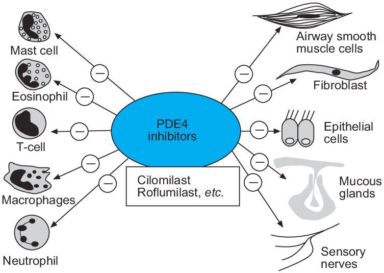
Phosphodiesterase-4 (PDE4) inhibitors have the potential to suppress inflammatory cells and structural cells in chronic obstructive pulmonary disease patients, giving a broad spectrum anti-inflammatory profile.
��
��
The cAMP Pathway as Therapeutic Target in Autoimmune and Inflammatory Diseases
Department of Dermatology, University Medical Center Mainz, Johannes Gutenberg-University Mainz, Mainz, Germany��
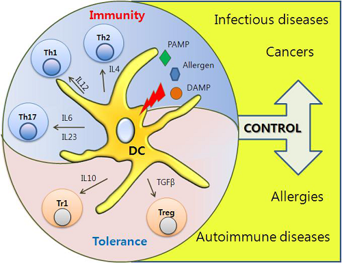
Dendritic Cells
As professional antigen-presenting cells of the immune system, DCs are equipped with a unique capability to induce and regulate adaptive immune responses. In DC, cyclic AMP suppresses the release of pro-inflammatory mediators (TNF-��, IL-17, IFN-��) (61) and promotes the release of anti-inflammatory mediators, such as IL-10 (62). As a functional consequence, cAMP concentrations in DC regulate T cell immunity (63). Pharmacological inhibition of cyclic nucleotide PDE4, which is highly expressed in DC, for example, suppresses the DC Th1-polarizing capacity (64, 65) and commands secretion of IL-6 and TGF-beta and subsequent induction of Th17 differentiation (66). It, thus, appears that cAMP levels differentially regulate cytokine production by DC as a response to changes in the microenvironment. Apart from spatio-temporal fine-tuning of DC activities, cAMP activities in DC depend on the stage of DC maturation: prostaglandin E2 (PGE2), a key inducer of cAMP, exerts a stimulatory function for immature DCs in peripheral tissues (67) but inhibitory function for mature DCs in lymph nodes (68).Frontiers | The cAMP Pathway as Therapeutic Target in Autoimmune and Inflammatory Diseases | Immunology
https://www.frontiersin.org/articles/10.3389/fimmu.2016.00123/full��
Figure 3. Calcium (Ca2+)�Ccalcineurin�Cnuclear factor of an activated T cell (NFAT) signaling pathway as a novel therapeutic target. Orai1 is a plasma membrane protein with four transmembrane segments. Stromal interaction molecule 1 (STIM1) is a single-pass transmembrane protein located in the endoplasmic reticulum (ER). The increase in intracellular Ca2+ concentration by Ca2+ entry through Ca2+ release-activated Ca2+ (CRAC)/Orai1 induces the phosphatase calcineurin. As a result, the activated calcineurin dephosphorylates several serine residues of the NFAT. The NFAT then translocates to the nucleus where it binds to DNA and regulates target gene expression. Some ions or small molecules, including La3+, SKF96365, and 2-ABP, are able to inhibit the CRAC channel. Again, already-commercialized cyclosporine A and tacrolimus inhibit calcineurin. Since dysregulated Ca2+ signaling is involved in the pathogenesis of autoimmune diseases, intervention of Ca2+ signaling in store-operated Ca2+ entry (SOCE) through the Orai1�CSTIM1 pathway may be a promising approach to control autoimmune diseases.
Calcineurin (CaN) is a calcium and calmodulin dependent serine/threonine protein phosphatase (also known as protein phosphatase 3, and calcium-dependent serine-threonine phosphatase).[2] It activates the T cells of the immune system and can be blocked by drugs. Calcineurin activates nuclear factor of activated T cell cytoplasmic (NFATc), a transcription factor, by dephosphorylating it. The activated NFATc is then translocated into the nucleus, where it upregulates the expression of interleukin 2 (IL-2), which, in turn, stimulates the growth and differentiation of the T cell response. Calcineurin is the target of a class of drugs called calcineurin inhibitors, which include ciclosporin, voclosporin, pimecrolimus and tacrolimus.Mechanism of action
When an antigen-presenting cell interacts with a T cell receptor on T cells, there is an increase in the cytoplasmic level of calcium, which activates calcineurin by binding a regulatory subunit and activating calmodulin binding.[3] Calcineurin induces transcription factors (NFATs) that are important in the transcription of IL-2 genes. IL-2 activates T-helper lymphocytes and induces the production of other cytokines. In this way, it governs the action of cytotoxic lymphocytes. The amount of IL-2 being produced by the T-helper cells is believed to influence the extent of the immune response significantly.�Ƶ�������ø(CaN)��һ�ָƺƵ�����������˿����/�հ��ᵰ������ø(Ҳ��Ϊ��������ø3����������˿����-�հ�������ø)�������Լ�������ϵͳ��Tϸ�������Ա�ҩ����ֹ���Ƶ�������øͨ��ȥ���ữ����Tϸ�����ʺ�����(NFATc)��һ��ת¼���ӡ������NFATc���ת�Ƶ�ϸ�����У��ϵ���ϸ������2 (IL-2)�ı�������̼�Tϸ���������ͷֻ���Ӧ���Ƶ�������ø��һ���Ϊ�Ƶ�������ø���Ƽ���ҩ��İе㣬����ҩ����������أ�voclosporin, pimecrolimus������Ī˾��
���û���
����ԭ�ʵ�ϸ����Tϸ���ϵ�Tϸ�����������ʱ��ϸ�����еĸ�ˮƽ�����ߣ��Ƶ�����øͨ��������ǻ���ϼ���Ƶ�����ϡ�[3]�Ƶ�������ø�յ�ת¼����(nfat)����IL-2�����ת¼������Ҫ���á�IL-2����t�����ܰ�ϸ�����յ�����ϸ�����ӵIJ�����ͨ�����ַ�ʽ����������ϸ�������ܰ�ϸ���Ļ��t����ϸ��������IL-2����������Ϊ������Ӱ������Ӧ��ij̶ȡ�Calcineurin - Wikipedia
https://en.wikipedia.org/wiki/Calcineurin[Magnesium: a natural calcium antagonist]
[Magnesium: a natural calcium antagonist] - PubMed
https://pubmed.ncbi.nlm.nih.gov/8926707/The dangers of magnesium deficiency | Dr. Thomas E. Levy ...
https://www.peakenergy.com/articles/nh20140407/The...
Apr 07, 2014 �� Probably the single most important property of magnesium in the body is its ability to act as a natural biological antagonist to calcium. As most adults have excess calciumthroughout their bodies, it is this reciprocal relationship between magnesium and calcium that makes most people in need of regular magnesium supplementation.��
Frontier Immunology, 10 March 2020 |
The Role of Calcium�CCalcineurin�CNFAT Signaling Pathway in Health and Autoimmune Diseases
Calcium (Ca2+) is an essential signaling molecule that controls a wide range of biological functions. In the immune system, calcium signals play a central role in a variety of cellular functions such as proliferation, differentiation, apoptosis, and numerous gene transcriptions. During an immune response, the engagement of T-cell and B-cell antigen receptors induces a decrease in the intracellular Ca2+ store and then activates store-operated Ca2+ entry (SOCE) to raise the intracellular Ca2+ concentration, which is mediated by the Ca2+ release-activated Ca2+ (CRAC) channels. Recently, identification of the two critical regulators of the CRAC channel, stromal interaction molecule (STIM) and Orai1, has broadened our understanding of the regulatory mechanisms of Ca2+ signaling in lymphocytes. Repetitive or prolonged increase in intracellular Ca2+ is required for the calcineurin-mediated dephosphorylation of the nuclear factor of an activated T cell (NFAT). Recent data indicate that Ca2+-calcineurin-NFAT1 to 4 pathways are dysregulated in autoimmune diseases. Therefore, calcineurin inhibitors, cyclosporine and tacrolimus, have been used for the treatment of such autoimmune diseases as systemic lupus erythematosus and rheumatoid arthritis. Here, we review the role of the Ca2+-calcineurin�CNFAT signaling pathway in health and diseases, focusing on the STIM and Orai1, and discuss the deregulated calcium-mediated calcineurin-NFAT pathway in autoimmune diseases.Psoriasis
Psoriasis is a chronic inflammatory skin disease characterized by various sized thick scaly erythematous plaques (108). The histopathology of psoriatic plaques shows epidermal proliferation and inflammation of the dermis (109). Both innate and adaptive immune cells, including keratinocytes and T cells, participate in the initiation and perpetuation of psoriasis (58, 110). Psoriasis is a well-established T cell-mediated skin disease (110, 111). In particular, various cytokines induce the activation of immune cells, particular Th1 and Th17 cells (111), and the functional imbalance of Th1 or Th17 over Tregs is considered a key pathway for the progression of psoriasis (111). For example, psoriatic skin lesions show a strong IFN-�� signature and have an abundance of IFN-�� (+) Th1 cells (112). An imbalance between Tregs and effector T (Teff) cells is observed in the peripheral blood of psoriasis patients (113). Moreover, the Tregs of psoriasis patients are functionally deficient in suppressing Teff cells (114). Recently, the association between IL-9 and the Th17 pathway has been reported in psoriasis. Expressions of IL-9 and IL-9R are markedly increased in psoriatic skin lesions (115), and IL-9 stimulates the production of IL-17A by CD4+ T cells isolated from patients with psoriasis (116).
It is well-established that calcineurin inhibitors suppress T cell activation and the differentiation of naive T cells to memory T cells (117). In particular, calcineurin inhibitors downregulate the expression of STAT1, IFN-��, and several IFN-��-downstream genes, repressing the generation of Th1 cells (118). Moreover, the expressions of IL-17, IL-22, and IL-17-inducible genes, including DEFB-2, LCN2, IL-1��, S100A12, and CCL20, are markedly suppressed by calcineurin inhibitors (119). Given the importance of Th1 and Th17 cells in psoriasis pathogenesis, the inhibition of the calcineurin�CNFAT pathway seems to be therapeutically relevant to psoriasis. Interestingly, the actions of calcineurin inhibitors are not limited to T cells. NFAT1 expression was first described by Northrop et al. (120) in mice skin, and then calcineurin expression was subsequently reported in the human epidermis (121). Calcineurin inhibitors reduce antigen presentation by Langerhans' cells and suppress neutrophil chemotaxis through the inhibition of psoriatic monocytes (122). Epidermal IL-1 and IL-8 expressions in psoriatic skin can be blocked by the calcineurin inhibitor cyclosporine (123). Indeed, cyclosporine and tacrolimus, both calcineurin inhibitors, have been widely used in psoriasis treatment with high efficacy (124).
STIM1 and Orai1 in keratinocytes, CRAC channels, have been implicated in the proliferation and differentiation of keratinocytes. It has been demonstrated that keratinocyte differentiation is induced by the change of extracellular Ca2+ concentration (125). Increased extracellular Ca2+ concentration triggers phospholipase C-mediated intracellular Ca2+ signals, which activate SOCE. Moreover, siRNA-mediated knockdown of either STIM1 or Orai1 suppresses SOCE and almost completely abolishes the Ca2+-mediated keratinocyte differentiation and growth (125). Menon and Elias (126) reported a defective Ca2+ gradient in the keratinocytes of psoriasis patients. Keratinocytes isolated from psoriasis patients showed a decreased response after Ca2+ store depletion as well as reduced mRNA/protein expression of CRAC channels (127, 128). In line with these findings, another study reported reduced mRNA and protein expression of TRPC channels (128), and the incubation of keratinocytes isolated from psoriasis patients with the TRPC6 agonist partly restores their differentiation and proliferation defect (129). Therefore, it remains to be determined whether Ca2+ sensing and signaling pathway plays an inductive or protective role in the pathologic differentiation and proliferation of keratinocytes in psoriasis.
Taken together, the earlier reports indicate that Ca2+ sensing and signaling pathway can be an excellent target for the treatment of psoriasis. Currently, phase 1 of a clinical trial of CRAC channel inhibitor for plaque psoriasis is in progress. CRAC channel inhibitors, a new class of oral immunomodulatory drugs, potently inhibit Orai1, Th1, Th2, and Th17-derived cytokine production and T cell proliferation, which are involved in chronic inflammatory responses in psoriasis (130).Frontiers | The Role of Calcium�CCalcineurin�CNFAT Signaling Pathway in Health and Autoimmune Diseases | Immunology
https://www.frontiersin.org/articles/10.3389/fimmu.2020.00195/full��
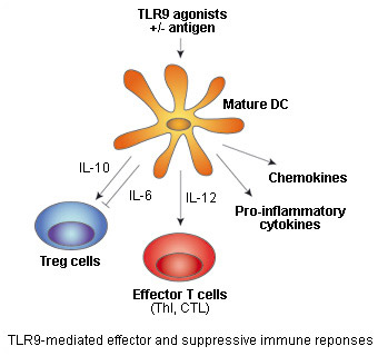
TLR9 agonists: double-edge sword for immune therapies
Toll-like receptor 9 (TLR9) senses unmethylated CpG dinucleotides, a hallmark of microbial DNA, that can be mimicked by synthetic oligonucleotides containing CpG motifs (CpG ODNs).
TLR9 stimulation by CpG DNA or CpG ODNs triggers intracellular signaling leading to the activation of macrophages, dendritic cells (DC) and B cells, and the production of cytokines, chemokines, and immunoglobulins. Subsequently, cytokines produced by DC, such as IL-12, induce the differentiation of naive T cells into T helper 1 (Th1) and cytotoxic T-cells (CTL).
CpG ODNs as vaccine adjuvants
Therefore, TLR9 agonists can elicit innate immune defenses and antigen T-cell specific responses, a property that underlines their development as vaccine adjuvants or immunotherapeutics for infectious diseases and cancer.
Studies in animal models have demonstrated that the immune defenses mounted by CpG ODNs alone or as vaccine adjuvants can protect against a variety of viral, bacterial, and parasitic diseases [1]. Promising results in the prophylactic treatment of hepatitis B have been obtained from phase III trials with a combination of a CpG ODN and hepatitis B surface antigen (Heplisav) [2].
CpG ODNs as antitumor agents
Antitumor activity of CpG ODNs has also been established in numerous mouse models. Encouraging results in the treatment of cancers have come from phase I and II clinical trials using CpG ODNs as a tumor vaccine adjuvant, monotherapy, or in combination with chemotherapy [2].
However, there have been also some disappointing results with one pharmaceutical company recently dropping its clinical program with a TLR9 agonist in non-small cell lung cancer. The interim data of two phase 3 trials of PF-3512676 (formerly called CpG 2006) showed that it failed to improve the clinical outcomes compared to chemotherapy alone [2].TLR9���弤����:�������Ƶ�˫�н�
toll������9 (TLR9)����δ������CpG�������ᣬ��������DNA��һ����־�����Ա�����CpG����ĺϳɹѺ�����(CpG ODNs)ģ�¡�
CpG DNA��CpG ODNs�̼�TLR9�ɴ���ϸ�����źţ����¾���ϸ������ͻ״ϸ��(DC)��Bϸ���ļ��������ϸ�����ӡ��������Ӻ������ס����DC������ϸ�����ӣ���IL-12���յ�ԭʼTϸ���ֻ�ΪT����1 (Th1)��ϸ������Tϸ��(CTL)��
CpG ODNs����������
��ˣ�TLR9�����������յ��������߷����Ϳ�ԭtϸ��������Ӧ����һ����ͻ����TLR9��Ϊ���������������Ʒ����ڸ�Ⱦ�Լ����Ͱ�֢�ķ�չ��
����ģ���о�����������ʹ��CpG ODNs����Ϊ���������������߷�������Ԥ�����ֲ�����ϸ���ͼ����漲��[1]������CpG ODN�������ױ��濹ԭ(Heplisav)[2]��III���ٴ�������Ԥ��������������ȡ������ϣ���Ľ����
CpG ODNs��Ϊ������ҩ��
CpG ODNs�Ŀ���������Ҳ��������С��ģ���еõ�֤ʵ��ʹ��CpG ODNs��Ϊ����������������ҩ���ƻ��뻯��[2]�������ư�֢��I�ں�II���ٴ�����ȡ�������˹���Ľ����
Ȼ����Ҳ��һЩ����ʧ���Ľ�������һ����ҩ��˾��������TLR9���������Ʒ�Сϸ���ΰ����ٴ���Ŀ������PF-3512676(ǰ��CpG 2006)�����ٴ����������������ʾ���뵥������[2]��ȣ�PF-3512676�����ܸ����ٴ���֡�
TLR9 agonists: double-edge sword for immune therapies
Toll-like receptor 9 (TLR9) senses unmethylated CpG dinucleotides, a hallmark of microbial DNA, that can be mimicked by synthetic oligonucleotides containing CpG motifs (CpG ODNs).
TLR9 stimulation by CpG DNA or CpG ODNs triggers intracellular signaling leading to the activation of macrophages, dendritic cells (DC) and B cells, and the production of cytokines, chemokines, and immunoglobulins. Subsequently, cytokines produced by DC, such as IL-12, induce the differentiation of naive T cells into T helper 1 (Th1) and cytotoxic T-cells (CTL).TLR9���弤����:�������Ƶ�˫�н�
toll������9 (TLR9)����δ������CpG�������ᣬ��������DNA��һ����־�����Ա�����CpG����ĺϳɹѺ�����(CpG ODNs)ģ�¡�
CpG DNA��CpG ODNs�̼�TLR9�ɴ���ϸ�����źţ����¾���ϸ������ͻ״ϸ��(DC)��Bϸ���ļ��������ϸ�����ӡ��������Ӻ������ס����DC������ϸ�����ӣ���IL-12���յ�ԭʼTϸ���ֻ�ΪT����1 (Th1)��ϸ������Tϸ��(CTL)��TLR9 and autoimmune diseases
Recent reports indicate that TLR9 may play a role in the pathogenesis of various autoimmune diseases, such as systemic lupus erythematosus (SLE). Under certain conditions,TLR9 is able to recognize self-DNA leading to the production of anti-DNA autoantibodies.
This discovery has prompted the development of specific inhibitors of TLR9. Paralleling the approach of stimulating TLR9 with CpG ODNs, it was found that suppressive ODNs exist that are able to inhibit TLR9 activation.The most potent inhibitory sequences contain TTAGGG multimers found in mammalian telomeres or a 5�� CCT, a C-free linker four to five bases long, and a GGG(G) tail. Some of these suppressive ODNs are able to inhibit an already ongoing immune response and therefore could be useful in the treatment of SLE [5].
This data illustrate the great potential of TLR9-based drugs for the treatment of infectious diseases, cancer and autoimmune diseases. However, as they can activate both the effector and suppressive arms of the immune system, more studies are needed to better understand the mechanisms involved allowing the development of safer and more effective TLR therapeutics.TLR9�����������Լ���
������о�������TLR9�����ڸ������������Լ����ķ��������з������ã���ϵͳ�Ժ���Ǵ�(SLE)����һ�������£�TLR9�ܹ�ʶ������dna���Ӷ�������dna�������塣
��һ���ִٽ���TLR9���������Ƽ��Ŀ�������CpG ODNs�̼�TLR9�ķ���ƽ�У��о����ִ����ܹ�����TLR9�����������ODNs������Ч���������а������鶯������з��ֵ�TTAGGG������5 ' CCT, 4��5��������IJ���c�������Ӻ�GGG(G)β�͡���Щ������odn�е�һЩ�ܹ������Ѿ����е����߷�Ӧ����˿��ܶ�SLE[5]���������á�
��Щ����˵���˻���tlr9��ҩ�������ƴ�Ⱦ������֢�����������Լ�������ľ�DZ����Ȼ�����������ǿ���ͬʱ��������ϵͳ��ЧӦ�ۺ����Ʊۣ���Ҫ������о������õ��˽��漰�Ļ��ƣ��Ӷ���������ȫ������Ч��TLR�Ʒ���https://www.invivogen.com/review-tlr9-agonists
��
��
��
��
Published 2009
Dendritic Cells as Danger-Recognizing Biosensors
Department of Bioscience and Biotechnology, College of Life Science, Sejong University, Seoul, 143-747, Korea
��
2.3.3. Autoimmune Diseases
Autoimmune diseases caused by autoreactive T cells or autoantibody-producing B cells can be initiated when self-antigens are presented by DCs. In addition, DCs in an autoimmune-prone condition can aggravate inflammatory immune responses by secreting IFN��/�� and TNF�� upon stimulation with necrotic cells caused by autoreactive T cells. TNF�� plays a critical role in the pathogenesis of rheumatoid arthritis and psoriasis, and DCs are the primary source of TNF�� [57]. Treatment of patients with TNF��-blocking therapeutic agents dramatically improved symptoms of arthritis and psoriasis. SLE is an autoimmune disease, characterized by the breakdown of immune tolerance by nuclear components and often gets worse by viral infection. Recently, it has been reported that pDCs are involved in the pathogenesis of SLE by secreting IFN��/�� [58]. pDCs, the main producer of IFN��/��, induce activation of autoreactive T cells and the production of autoreactive antibodies by making immature DCs become mature DCs [59]. It is now believed that pDCs mediate IFN��/�� production by signaling through TLR7 and TLR9, which recognize viral nucleic acid, or via DNA-binding proteins such as HMGB1 and RAGE [11,60].
2.3.4. Allergies
Although inhaled allergens are taken up by DCs and presented to T cells, tolerance is induced in healthy individuals. However, in allergic disease conditions, immune tolerance against inhaled allergens becomes broken, resulting in the presentation of allergens by DCs to T cells. Upon recognition of allergens, T cells differentiate toward Th2 cells, which give aid to B cells and produce allergen-specific immunoglobulin (Ig) E. Skin epithelial cells directly or indirectly influence DCs to induce allergic immune responses. Skin epithelial cells help DCs to sample allergens infiltrated into skin and also regulate cytokine production of DCs to help differentiation of naïve CD4+ T cells into Th2 cells. Thymic stromal lymphopoietin or granulocyte macrophage colony stimulating factor (GM-CSF), produced by epithelial cells, has been known to induce Th2 polarization [61,62]. In addition, it has been recently reported that inhaled allergens with enzymatic activity act on DCs to induce Th2 differentiation [62].
4. Conclusions
DCs play a critical role in determining the polarization of T cell immune responses, which have a direct impact on what types of antibody isotypes B cells produce. In other words, DCs can direct which adaptive immune responses are induced. Research on the functions of DCs should become a central focus of the immunology field because DCs are involved in various diseases, including infectious diseases, cancer, autoimmune diseases, and allergies.
Through recognition of appropriate danger signals, DCs help to protect the host from a variety of diseases and maintain a micro-environment for optimal immunity in healthy individuals. If DCs do not detect danger signals, the host immune system does not induce immune responses against pathogens and also may not induce immune tolerance upon induction of autoimmune responses. Considering the importance of DC functions in immunological regulation, DCs have been the focus for therapeutics for many diseases including cancers. Here, we have introduced the cytokine reporter mouse model as a tool for monitoring activation of DCs. To achieve better understanding of DC functions, it will be important to expand the studies of DC subsets in healthy and diseased states. To conclude, we anticipate a dramatic increase in the number of studies on DCs that will be based on populations that are isolated from mouse and human lymphoid organs or are cultured in vitro using DC growth factors.2.3.3�����������Լ���
��DCs�ʽ�������ԭʱ����������Ӧ��Tϸ����������������Bϸ����������������Լ����ͻᷢ�������⣬�������������������£���ͻ״ϸ��ͨ��������Ӧ��Tϸ������Ļ���ϸ���̼�����IFN��/�º�TNF�����ɼ�����֢���߷�Ӧ��TNF�������ʪ�ؽ�����м���ķ��������з�����Ҫ���ã�DCs��TNF��[57]����Ҫ��Դ��TNF���������ҩ���������ƹؽ�����м��֢״��SLE��һ�����������Լ�������������ͨ���˳ɷ��ƻ��������ܣ�����������Ⱦ��������б�����pDCsͨ������IFN��/��[58]����SLE�ķ������ơ�pDCs��IFN��/�µ���Ҫ�����ߣ�ͨ��ʹδ�������ͻ״ϸ����ɳ������ͻ״ϸ��[59]���յ�������Ӧ��Tϸ���Ļ��������Ӧ�Կ���IJ�����������ΪpDCsͨ��ʶ�������TLR7��TLR9�ź�ת������ͨ��HMGB1��RAGE��dna��ϵ��鵼IFN��/�µIJ���[11,60]��
2.3.4������
��Ȼ����Ĺ���ԭ����ͻ״ϸ�����ղ��ʵݸ�Tϸ�������ڽ��������п��յ����ܡ�Ȼ�����ڹ����Լ����������£��������������Ӧԭ�����ƣ�����DCs����Ӧԭ�ύ��Tϸ����Tϸ����ʶ�����ԭ����Th2ϸ���ֻ���Th2ϸ������Bϸ����������ԭ������������(Ig) E.Ƥ����Ƥϸ��ֱ�ӻ���Ӱ��dc�յ���������Ӧ��Ƥ����Ƥϸ��������ͻ״ϸ��������Ƥ���ı�Ӧԭ����ȡ������������ͻ״ϸ����ϸ����������������ԭʼCD4+ Tϸ���ֻ�ΪTh2ϸ��������Ƥϸ�����������ٻ����ܰ�ϸ�������ڻ���ϸ������ϸ������̼�����(GM-CSF)�ѱ�֤ʵ���յ�Th2����[61,2]�����⣬����б����ƣ�����ľ���ø���Եı�Ӧԭ������DCs���յ�Th2�ֻ�[62]��
4. ����
DCs�ھ���Tϸ�����߷�Ӧ�ļ�������������������Ҫ�����ã���ֱ��Ӱ��Bϸ�������������͵Ŀ���ͬ�͡����仰˵����ͻ״ϸ������ָ����Щ��Ӧ�����߷�Ӧ���յ�������DCs�漰���ּ�����������Ⱦ������֢�����������Լ����������ȣ���˶�DCs���ܵ��о�Ӧ��Ϊ����ѧ������ص㡣
ͨ��ʶ���ʵ���Σ���źţ�DCs�����ڱ����������ܸ��ּ�������Ϯ����ά�ֽ�������������ߵ����������DCsû�м�Σ���źţ���������ϵͳ�Ͳ����յ��Բ�ԭ�������Ӧ��Ҳ���ܲ����յ���������Ӧ�����������ܡ�����DC���������ߵ����е���Ҫ�ԣ�DC�ѳ�Ϊ������֢���ڵ����༲�����Ƶ��ص㡣��������ǽ�����ϸ�����ӱ���С��ģ����Ϊ���DCs����Ĺ��ߡ�Ϊ�˸��õ�����ֱ����Ĺ��ܣ������ͼ���״̬�µ�ֱ������Ⱥ���о�������Ҫ�ġ���������������Ԥ��DC�о����������������ӣ���Щ�о������ڴ�С������ܰ����ٷ����Ⱥ�壬��ʹ��DC������������������Ⱥ�塣http://www.mdpi.com/1424-8220/9/9/6730/htm
��
Cells, Mar 14, 2020
Glycogen Metabolism Supports Early Glycolytic Reprogramming and Activation in Dendritic Cells in Response to Both TLR and Syk-Dependent CLR Agonists
Dendritic cells (DCs) increase their metabolic dependence on glucose and glycolysis to support their maturation, activation-associated cytokine production, and T-cell stimulatory capacity. We have previously shown that this increase in glucose metabolism can be initiated by both Toll-like receptor (TLR) and C-type lectin receptor (CLR) agonists. In addition, we have shown that the TLR-dependent demand for glucose is partially satisfied by intracellular glycogen stores. However, the role of glycogen metabolism in supporting CLR-dependent DC glycolytic demand has not been formally demonstrated. In this work, we have shown that DCs activated with fungal-associated ��-glucan ligands exhibit acute glycolysis induction that is dependent on glycogen metabolism. Furthermore, glycogen metabolism supports DC maturation, inflammatory cytokine production, and priming of the nucleotide-binding domain, leucine-rich-containing family, pyrin domain-containing-3 (NLRP3) inflammasome in response to both TLR- and CLR-mediated activation. These data support a model in which different classes of innate immune receptors functionally converge in their requirement for glycogen-dependent glycolysis to metabolically support early DC activation. These studies provide new insight into how DC immune effector function is metabolically regulated in response to diverse inflammatory stimuli.��ԭ��л֧�������ǽͽ��ر�̺���ͻ״ϸ���Ļ��ͬʱ��ӦTLR��syk������CLR������
��ͻ״ϸ��(dc)���Ӷ������Ǻ��ǽͽ�Ĵ�л��������֧������졢�������ϸ�����ӵIJ�����tϸ���Ĵ̼�����������֮ǰ�Ѿ�֤�������������Ǵ�л�����ӿ�����toll������(TLR)��c������������(CLR)���������������⣬���Ƿ�������tlr������������ͨ��ϸ������ԭ�洢�õ����㡣Ȼ������ԭ��л��֧��clr������DC�ǽͽ������е�������δ�õ���ʽ֤������������У������Ѿ�֤���˱������ئ�-�Ͼ������弤�����ͻ״ϸ�����ֳ���������ԭ��л�ļ����ǽͽ��յ������⣬��ԭ��л֧����TLR��clr�鵼�ļ����£�DC���죬��֢ϸ�����ӵIJ��������ս������������ļ��壬pyrin�������-3 (NLRP3)����С�����������Щ����֧����һ��ģ�ͣ������ģ���У���ͬ���������������������ԭ�������ǽͽ������й�����ͬ���Դ�л��ʽ֧������DC�����Щ�о�ΪDC����ЧӦ�����ڲ�ͬ��֢�̼��µĴ�л�����ṩ���µļ��⡣
Keywords: dendritic cells; glucose; glycogen; glycolysis; innate immunity; metabolism.Glycogen Metabolism Supports Early Glycolytic Reprogramming and Activation in Dendritic Cells in Response to Both TLR and Syk-Dependent CLR Agonists - PubMed
https://pubmed.ncbi.nlm.nih.gov/32183271/#:~:text=Dendritic%20cells%20%28DCs%29%20increase%20their%20metabolic%20dependence%20on,receptor%20%28TLR%29%20and%20C-type%20lectin%20receptor%20%28CLR%29%20agonists.��
��
Metabolic Control of Dendritic Cell Functions: Digesting Information
Immunobiology Laboratory, Centro Nacional de Investigaciones Cardiovasculares (CNIC), Madrid, Spain��
Metabolic Control of Dendritic Cell Development
Natural dendritic cells (DCs) present in steady state comprise type 1 conventional DCs (cDC1s), type 2 cDCs (cDC2s), double negative (CD8/CD103�C CD11b�C) DCs (DN-DCs), and plasmacytoid DCs (pDCs; Table 1). Natural DCs derive from myeloid progenitors in the bone marrow and require FMS-like tyrosine kinase 3 ligand (FLT3L) to differentiate via the common DC progenitor (CDP) and DC precursors (pre-DCs). In addition, other cells that are functionally similar to DCs, such as Langerhans cells (LCs), can derive from embryonic precursors. Moreover, during inflammatory settings, DCs can develop from blood monocytes (moDCs; Table 1).��
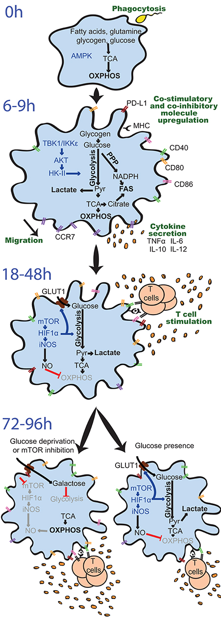
Figure 3. Differential regulation and effects of glycolysis induction in GM-DCs upon stimulation over time. Resting GM-DCs (top) display a basal metabolism with active AMPK (AMP-activated protein kinase) and fatty acids, glutamine, glycogen, and glucose being fully oxidized to generate energy by oxidative phosphorylation (OXPHOS). Upon early stimulation after 6�C9 h, GM-DCs are activated and exhibit transiently enhanced OXPHOS/mitochondrial membrane potential and an increased glycolytic metabolism mainly using glucose from intracellular glycogen stores. The induction of glycolysis is predominantly driven by a TBK1-IKK��/AKT/HK-II axis and largely devoted to fatty acid synthesis (FAS). Moreover, enhanced early glycolytic activity of GM-DCs is vital for their migration and upregulation of co-stimulatory/inhibitory molecules as well as cytokines. At later time points about 18�C48 h after robust stimulation, a mTOR/HIF1��/iNOS axis is activated in GM-DCs, leading to enforced glycolysis via upregulation of glucose importers such as GLUT1 and inhibition of OXPHOS via nitric oxide (NO). This fostered glycolytic activity appears crucial for the interaction of GM-DCs with T cells. Nevertheless, the sustained inhibition of OXPHOS by NO and reliance on glycolysis for energy generation can reduce the ability of GM-DCs to stimulate T cells in the long term. Glucose deprivation or mTOR inhibition can preserve metabolic flexibility and functional OXPHOS in GM-DCs, sustaining their activity at least during 72�C96 h and extending their life span.
AKT, protein kinase B; CCR7, C-C chemokine receptor type 7; CD, cluster of differentiation; GLUT1, glucose transporter 1; GM-DC, GM-CSF, mouse GM-CSF-induced DCs; HIF1��, hypoxia-inducible factor 1-alpha; HK-II, hexokinase II; IKK��, IkB kinase; IL, interleukin; iNOS, inducible nitric oxide synthase; MHC, major histocompatibility complex; mTOR, mammalian target of rapamycin; NADPH, nicotinamide adenine dinucleotide phosphate; PD-L1, programmed death-ligand 1, Pyr, pyruvate; PPP, pentose phosphate pathway; TBK1, TANK-binding kinase 1; TCA, Tricarboxcylic acid cycle; TNF��, tumor necrosis factor ��.��
Requirement of Glycolysis for Functions of Activated Dendritic Cells
Interrupting the glucose-to-pyruvate pathway significantly impairs DC maturation, upregulation of co-stimulatory molecules, cytokine secretion, and T cell stimulatory capacity in the long term (Figure 3). For example, pharmacological blockade of glycolysis using 2-deoxyglucose (2-DG), genetic deficiency of glycolytic enzymes such as ��-enolase (ENO1), or overexpression of lactate dehydrogenase A (LDHA) or pyruvate dehydrogenase kinase 1 (PDK1) (Figure 2) prevents GM-DC maturation and immunogenicity upon stimulation with LPS or Chlamydia (13, 47, 49, 57) and can skew GM-DCs toward inducing Th17 and regulatory T cells (Treg) rather than Th1 and Th2 responses (49). In line, natural mouse cDC1s and cDC2s isolated from the spleen decrease expression of co-stimulatory molecules, IL-12 production, and activation of CD4+ and CD8+ T cells when activated by LPS in the presence of 2-DG (49). pRNA-stimulated human blood cDC2s require glycolytic activity for activation, evidenced by TNF�� production, CD86, and programmed death ligand 1 (PD-L1) expression (53). Treatment of primary human pDCs with 2-DG upon influenza A virus stimulation also reduces co-stimulatory molecule and type I interferon (IFN-I) expression (52), while another study rather suggests induction of glutamine-fueled OXPHOS upon pRNA stimulation of human blood pDCs (53). However, the effects of inhibition of glycolysis by 2-DG in DCs have to be taken with caution, as 2-DG itself deregulates cytokine expression of human moDCs in vitro by activation of the endoplasmic reticulum (ER) stress response via the sensor inositol-requiring protein 1�� (IRE1��) (50). In addition, 2-DG can impair the TCA cycle, OXPHOS, and ATP levels, as recently described in macrophages (58).
Other DC functions such as phagocytosis do not seem to be affected by inhibition of glycolysis during stimulation of human moDCs (50). However, reduced endocytic/phagocytic activity in aging mouse spleen cDC1s and DN-DCs [termed merocytic DCs (mcDCs)] and a resulting decline in antigen cross-presentation are linked to mitochondrial dysfunction with decreased basal OCR and ����m as well as enhanced proton leakage and ROS. Importantly, inhibition of ATP synthase by oligomycin or the uncoupling agent carbonyl cyanide 4-(trifluromethoxy)phenyl-hydrazone (FCCP) corroborates the diminished phagocytosis of cDC1s and DN-DCs/mcDCs (22). Moreover, antigen uptake seems to decrease in GM-DCs in hypoxia, when glycolytic activity is increased by HIF1�� stabilization, which is also observed in human moDCs after stimulation (47, 50).
In contrast, glucose and enhanced glycolytic activity are required for the ability of DCs to migrate (Figure 3). Independently of stimulation, glucose-deprived GM-DCs show reduced mobility, increased rounded morphology losing dendrites, and impaired oligomerization of CCR7, the chemokine receptor driving DC migration toward LNs. Subsequently, glucose limitation or 2-DG presence prevents migration of GM-DCs as well as splenic CD11c+ cDCs both in vitro and in vivo (49, 56). In line, HIF1��-deficient GM-DCs, which largely fail to induce glycolysis (see the section Sustained Glycolysis: The Role of HIF1��), display reduced CCR7 levels, and GM-DCs differentiated in hypoxic conditions exhibit elevated migratory potential in vitro and in vivo that is dependent on HIF1�� (14).
Overall, early induction of glycolysis emerges as a general feature of immunogenic activation of most cultured DCs and primary DC subsets and appears necessary for several aspects of their maturation such as upregulation of co-stimulatory surface molecules and cytokine production, despite having no major effects on phagocytosis or antigen uptake. However, DC activation leads to cytoskeletal changes that support increased migratory capacity to migrate toward LNs and T cell zones, which is also affected by early induced glycolysis. Ultimately, in light of those findings, glycolytic increase in DCs upon stimulation is vital for adequate induction of adaptive T cell responses (59) and, hence, regulates immune homeostasis (Figure 3).
https://www.frontiersin.org/articles/10.3389/fimmu.2019.00775/full��
Significance of DC-LAMP and DC-SIGN expression in psoriasis vulgaris lesions
In psoriasis vulgaris lesions, dendritic cells are massively recruited to the dermis, where these cells mature gradually and express a large amount of DC-LAMP and DC-SIGN, thereby activating T lymphocytes and participating in the process of immunological inflammation. The high expression levels of DC-LAMP and DC-SIGN are positively correlated.
Author: Ma Wei-yuan, Liu Wen-ting, Zhao Chen, Sun Qing
Cited by: 10
Publish Year: 2011
Significance of DC-LAMP and DC-SIGN expression in psoriasis vulgaris lesions - ScienceDirect
https://www.sciencedirect.com/science/article/abs/pii/S0014480011000554#:~:text=In%20psoriasis%20vulgaris%20lesions%2C%20dendritic%20cells%20are%20massively,levels%20of%20DC-LAMP%20and%20DC-SIGN%20are%20positively%20correlated.
��Front. Immunol., 19 June 2018
T Cell Hierarchy in the Pathogenesis of Psoriasis and Associated Cardiovascular Comorbidities
The key role of T cells in the pathogenesis of cutaneous psoriasis has been well described in the last decade and the knowledge of the relative role of the different subsets of T cells in psoriasis pathogenesis has considerably evolved. Now, it is clear that IL-17A-producing T cells, including Th17/Tc17, have a central role in the pathogenesis of cutaneous psoriasis and therapies blocking the IL-17A pathway show high clinical efficacy. By contrast, the contribution of IFN��-producing T cells has progressively become less clear because of the lack of efficacy of anti-IFN�� antibodies in clinical studies. In parallel, the role of CD8+ T cells specific for self-antigens has been revived and increasing evidence now indicates that in psoriatic skin the majority CD8+ T cells are present in the form of epidermal tissue-resident memory T cells. In the last years it also emerged the possibility of a contribution of T cell recirculation in the pathogenesis of psoriasis and its systemic manifestations.ǰ�档Immunol��2018��6��19��
Tϸ���ȼ�����м���������ƺ������Ѫ�ܹ����е�����
Tϸ����Ƥ����м�����������еĹؼ������ڹ�ȥʮ���еõ��˺ܺõ����������Ҷ�Tϸ����ͬ��Ⱥ����м�����������е�������������൱��Ľ�չ�������Ѿ���ȷIL-17A����Tϸ��������Th17/Tc17����Ƥ����м���ķ������������������ã����IL-17Aͨ·��������ʾ���ϸߵ��ٴ���Ч�����֮�£������ٴ��о���ȱ����IFN�ÿ������Ч�ԣ�IFN�ò���Tϸ������������ò���������ͬʱ�����������ԭ��CD8+ Tϸ��������Ҳ�õ��˻ָ���Խ��Խ���֤�ݱ���������м��Ƥ���У������CD8+ Tϸ���Ա�Ƥ��֯פ������Tϸ������ʽ���ڡ���������Tϸ��ѭ������м���������Ƽ���ȫ�������з������õĿ�����Ҳ�����ˡ�Frontiers | T Cell Hierarchy in the Pathogenesis of Psoriasis and Associated Cardiovascular Comorbidities | Immunology
https://www.frontiersin.org/articles/10.3389/fimmu.2018.01390/full��
Biomedicine & Pharmacotherapy
Volume 131, November 2020,
Luteolin attenuates imiquimod�Cinduced psoriasis-like skin lesions in BALB/c mice via suppression of inflammation response
School of Biomedical and Pharmaceutical Sciences, Guangdong University of Technology, 100 Waihuanxi Road, Guangzhou, 510006, PR China
b
Guangdong Provincial Key Laboratory of Emergency Test for Dangerous Chemicals, Guangdong Institute of Analysis, Guangzhou, 510070, PR China
Highlights
•
Luteolin ameliorated psoriasis-like skin lesions in an imiquimod-induced mouse model.
•
Luteolin suppressed the cutaneous macrophage infiltration and cytokine release in vivo.
•
Luteolin attenuated inflammatory response in a macrophage cell line.
Abstract
Psoriasis is considered as a common chronic immune-mediated skin disorder characterized by abnormal keratinocyte proliferation. Luteolin, an anti-inflammatory natural flavonoid with well-accepted inhibition effect against keratinocyte proliferation, was hypothesized to have a potential therapeutic effect for psoriasis. In this paper, we investigated the relieving effect of luteolin against imiquimod-induced psoriasis-like lesions on BALB/c mice and its possible anti-inflammatory mechanisms in lipopolysaccharide-stimulated macrophages (RAW264.7 cells). We found that luteolin ameliorated psoriasis-like skin lesions, suppressed the cutaneous infiltration of macrophages, T cells and neutrophils, and downregulated the expression of cytokines like IL-6, IL-1��, TNF-��, IL-17A and IL-23 in both skin lesions and eyeball blood of model mice. In vitro, we observed luteolin significantly suppressed the levels of psoriasis-related pro-inflammatory cytokines, such as IL-17A, IL-6, TNF-�� and IL-23, and inflammatory mediators like nitric oxide NO, inducible NOS, COX-2 in RAW264.7 cells. The anti-inflammatory activity was accomplished by inhibiting NF-��B expression and activation. This study demonstrates luteolin is effective in alleviating psoriasis-like skin lesions and downregulating inflammatory response via NF-��B pathway, suggesting luteolin as a potential molecule for further therapeutic research of inflammation-related skin diseases like psoriasis.
ľϬ����(luteolin)��ͨ��������֢��Ӧ�������Ī���յ���BALB/cС����м����Ƥ��
ͻ����
• ľϬ���ظ������Ī���յ�С��ģ������м����Ƥ�����ˡ�
• ľϬ��������������Ƥ������ϸ�������ϸ�������ͷš�
• ľϬ���ؿɼ�������ϸ������֢��Ӧ��
��м������Ϊ��һ�ֳ������������߽鵼��Ƥ�����������쳣�Ľ���ϸ����ֳΪ������ľϬ������һ����Ȼ�Ŀ������ͪ���Խ���ϸ����ֳ�����õ��������ã�����Ϊ����м����DZ�ڵ��������á������о�ľ���ض�֬���Ǵ̼�����ϸ��(RAW264.7ϸ��)�����Ī���յ�����м����Ƥ��Ļ������ü�����ܵĿ����ơ����Ƿ���ľϬ���ؿ��Ը�����м����Ƥ�����ƾ���ϸ����Tϸ����������ϸ����Ƥ�������µ�ģ��С��Ƥ�����Ѫ��IL-6��IL-1�¡�TNF-����IL-17A��IL-23��ϸ�����ӵı��������ʵ���У�ľϬ��������������RAW264.7ϸ��������м����صĴ�������IL-17A��IL-6��TNF-����IL-23��ˮƽ���Լ���֢����NO���յ���NOS��COX-2��ˮƽ���俹������ͨ������NF-��B�ı���ͻ��ʵ�ֵġ����о�����ľϬ���ؿ�ͨ��NF-��Bͨ·������м����Ƥ���µ���֢��Ӧ����ʾľϬ����������Ϊ��м������֢���Ƥ�����Ľ�һ�������о��ķ��ӡ�
Luteolin attenuates imiquimod�Cinduced psoriasis-like skin lesions in BALB/c mice via suppression of inflammation response - ScienceDirect
https://www.sciencedirect.com/science/article/pii/S0753332220308891#:~:text=Macrophages%20were%20found%20to%20accumulate%20in%20psoriatic%20skin,keratinocytes%20and%20the%20skin%20inflammation%20observed%20in%20psoriasis.��
Cytokine Pathways in Psoriasis and Psoriatic Arthritis
Psoriatic disease is a systemic autoimmune disease mostly associated with skin and joint involvement (psoriatic arthritis). Strong evidences from clinical studies and experimental models suggest that both innate and adaptive immune responses are involved in its pathogenesis. Psoriatic disease used to be regarded as a Th1-driven disease, now there are substantial evidences to suggest regulatory role of Th17 cells as well in the pathogenesis of psoriasis and psoriatic arthritis. Cytokines play a critical role; besides IFN-�� and TNF��, IL-23/Th17 pathway plays a dominant role in the inflammatory and proliferative cascades of both the skin and joint tissues. Recently in a series of elegant experiments using mouse models and human tissues it has been demonstrated that IL-23 induced Th17 cytokines (IL-17 and IL-22) can contribute to all four pathologic events in a psoriatic disease: development of psoriatic plaque, pannus formation in the joint, joint erosion and new bone formation.
����м������м���ؽ����е�ϸ������ͨ·
��м����һ��ȫ�����������Լ���������Ƥ���ؽ��������(��м���ؽ���)���ٴ��о���ʵ��ģ�͵�����֤�ݱ����������Ժ���Ӧ�����߷�Ӧ�������䷢�����ơ���м��������Ϊ��th1�����ļ����������д���֤�ݱ���Th17ϸ������м������м���ؽ��ķ���������Ҳ���ӵ������á�ϸ��������ؼ�����;����IFN-�ú�TNF���⣬IL-23/Th17ͨ·��Ƥ���ؽ���֯����֢����ֳ���������������á������һϵ������ʵ����С��ģ�ͺ�������֯�Ѿ�֤��IL-23��ӦTh17ϸ������(IL-17��il - 22����)����Ϊ�����ĸ������¼�ţƤѢ����:��м���߿�ķ�չ,Ѫ�����γ�����,��ͬ��ʴ���¹��γɡ�
Cytokine Pathways in Psoriasis and Psoriatic Arthritis | SpringerLink
https://link.springer.com/chapter/10.1007/978-3-319-19530-8_9��
Frontier in Immunology Oct 6, 2020
Lactate Suppresses Macrophage Pro-Inflammatory Response to LPS Stimulation by Inhibition of YAP and NF-��B Activation via GPR81-Mediated Signaling
Kun Yang1,2†, Jingjing Xu1†‡, Min Fan1,2, Fei Tu1,2, Xiaohui Wang1, Tuanzhu Ha1,2, David L. Williams1,2 and Chuanfu Li1,2*
1Department of Surgery, Quillen College of Medicine, East Tennessee State University, Johnson City, TN, United States
2Center of Excellence for Inflammation, Infectious Disease and Immunity, Quillen College of Medicine, East Tennessee State University, Johnson City, TN, United StatesRecent evidence from cancer research indicates that lactate exerts a suppressive effect on innate immune responses in cancer. This study investigated the mechanisms by which lactate suppresses macrophage pro-inflammatory responses. Macrophages [Raw 264.7 and bone marrow derived macrophages (BMDMs)] were treated with LPS in the presence or absence of lactate. Pro-inflammatory cytokines, NF-��B and YAP activation and nuclear translocation were examined. Our results show that lactate significantly attenuates LPS stimulated macrophage TNF-�� and IL-6 production. Lactate also suppresses LPS stimulated macrophage NF-��B and YAP activation and nuclear translocation in macrophages. Interestingly, YAP activation and nuclear translocation are required for LPS stimulated macrophage NF-��B activation and TNF�� production. Importantly, lactate suppressed YAP activation and nuclear translocation is mediated by GPR81 dependent AMKP and LATS activation which phosphorylates YAP, resulting in YAP inactivation. Finally, we demonstrated that LPS stimulation induces an interaction between YAP and NF-��B subunit p65, while lactate decreases the interaction of YAP and NF-��B, thus suppressing LPS induced pro-inflammatory cytokine production. Our study demonstrates that lactate exerts a previously unknown role in the suppression of macrophage pro-inflammatory cytokine production via GPR81 mediated YAP inactivation, resulting in disruption of YAP and NF-��B interaction and nuclear translocation in macrophages.
����ͨ��gpr81�鵼���ź�ͨ·����YAP��NF-��B����Ӷ����ƾ���ϸ����LPS�̼��Ĵ���֢��Ӧ
�����֢�о���֤�ݱ����������֢���������߷�Ӧ���������á����о�̽�������������ƾ���ϸ������Ӧ�Ļ��ơ�����ϸ��[Raw 264.7������Դ����ϸ��(bone marrow derived Macrophages, BMDMs)]��������ڻ��ڵ�����±�LPS������������ϸ�����ӡ�NF-��B��YAP�����תλ�����ǵ��о�������������������������LPS�̼��ľ���ϸ��TNF-����IL-6�IJ��������ỹ��������LPS�̼��ľ���ϸ��NF-��B��YAP�Ļ�Լ�����ϸ���ĺ�תλ����Ȥ���ǣ�LPS�̼��ľ���ϸ��NF-��B�����TNF -��������ҪYAP����ͺ�תλ����Ҫ���ǣ���������YAP����ͺ�תλ����GPR81������AMKP��LATS����鵼�ģ���Щ����ʹYAP���ữ������YAPʧ�������Ƿ���LPS�̼��յ�YAP��NF-��B�ǻ�p65����ã������ή��YAP��NF-��B����ã��Ӷ�����LPS�յ��Ĵ���֢ϸ�����ӵIJ��������ǵ��о�����������ͨ��GPR81�鵼��YAPʧ�������ƾ���ϸ��������ϸ�����Ӳ����з�������ǰδ֪�����ã����¾���ϸ����YAP��NF-��B����õ��ж��Լ���תλ��
Frontiers | Lactate Suppresses Macrophage Pro-Inflammatory Response to LPS Stimulation by Inhibition of YAP and NF-��B Activation via GPR81-Mediated Signaling | Immunology
https://www.frontiersin.org/articles/10.3389/fimmu.2020.587913/full��
Lactic acid delays the inflammatory response of human monocytes
a Department of Internal Medicine III, University Hospital Regensburg, Franz-Josef-Strauß-Allee 11, 93053 Regensburg, Germany
b RCI Regensburg Center for Interventional Immunology, University Hospital Regensburg, Franz-Josef-Strauß-Allee 11, 93053 Regensburg, Germany
Received 20 December 2014, Available online 9 January 2015.
Highlights
• Lactic acid broadly delays LPS-induced gene expression in human monocytes.
• Expression of important monocyte effector molecules is affected by lactic acid.
• Interference of lactic acid with TLR signaling causes the delayed gene expression.
•The profound effect of lactic acid might contribute to immune suppression in tumors.
Abstract
Lactic acid (LA) accumulates under inflammatory conditions, e.g. in wounds or tumors, and influences local immune cell functions. We previously noted inhibitory effects of LA on glycolysis and TNF secretion of human LPS-stimulated monocytes. Here, we globally analyze the influence of LA on gene expression during monocyte activation. To separate LA-specific from lactate- or pH-effects, monocytes were treated for one or four hours with LPS in the presence of physiological concentrations of LA, sodium lactate (NaL) or acidic pH. Analyses of global gene expression profiles revealed striking effects of LA during the early stimulation phase. Up-regulation of most LPS-induced genes was significantly delayed in the presence of LA, while this inhibitory effect was attenuated in acidified samples and not detected after incubation with NaL. LA targets included genes encoding for important monocyte effector proteins like cytokines (e.g. TNF and IL-23) or chemokines (e.g. CCL2 and CCL7). LA effects were validated for several targets by quantitative RT-PCR and/or ELISA. Further analysis of LPS-signaling pathways revealed that LA delayed the phosphorylation of protein kinase B (AKT) as well as the degradation of I��B��. Consistently, the LPS-induced nuclear accumulation of NF��B was also diminished in response to LA. These results indicate that the broad effect of LA on gene expression and function of human monocytes is at least partially caused by its interference with immediate signal transduction events after activation. This mechanism might contribute to monocyte suppression in the tumor environment.�����ӻ������ϸ������֢��Ӧ
ͻ����
•����㷺�ӳ�lps�յ��������ϸ��������
•��Ҫ����ϸ��ЧӦ���ӵı����ܵ������Ӱ�졣
•�����TLR�źŵĸ��ŵ��»�������ӳ١�
•�������ԶӰ������������������������ơ�
ժҪ
����(LA)����֢�����»��ۣ������˿ڻ������У���Ӱ��ֲ�����ϸ�����ܡ�������ǰע�LA��lps�̼����˵���ϸ�����ǽͽ��TNF���ڵ��������á����������ȫ�����LA�ڵ���ϸ����������жԻ�������Ӱ�졣Ϊ�˽�LA�������������phЧӦ���ֿ�����������Ũ��ΪLA��������(NaL)������ph������£�����ϸ����LPS����1��4Сʱ���������������ķ�����ʾ�������ڴ̼��Σ�LA�������������á���LA���ڵ�����£������lps�յ�������ϵ��������ӳ٣������������������ữ��Ʒ�м���������NaL������û�м���LA�İб����������Ҫ����ϸ��ЧӦ���Ļ�����ϸ������(��TNF��IL-23)����������(��CCL2��CCL7)��ͨ��RT-PCR��/��ELISA������֤LA�Զ���е�����á���һ������lps�ź�ͨ·����LA�ӻ��˵���øB (AKT)�����ữ�Լ�I��B���Ľ��⡣ͬ����lps�յ���NF -��B�ĺ˻���Ҳ��LA�������¼��١���Щ���������LA���˵���ϸ�����������ܵĹ㷺Ӱ�����ٲ������������ڼ����ֱ�Ӹ����ź�ת���¼����¡���һ���ƿ��������ڵ���ϸ�������������е����ơ�
Lactic acid delays the inflammatory response of human monocytes - ScienceDirect
https://www.sciencedirect.com/science/article/abs/pii/S0006291X1500011X��
IMMUNOBIOLOGY| MARCH 1, 2006
Tumor-derived lactic acid modulates dendritic cell activation and antigen expressionThe Department of Hematology and Oncology, Institute of Pathology, University of Regensburg; and the Institute of Physiology and Pathophysiology, University of Mainz, Germany.
The tumor milieu can influence dendritic cell (DC) differentiation. We analyzed DC differentiation in a 3-dimensional tumor model and propose a new mechanism of DC modulation by the tumor environment. Monocytes were cultured in the presence of IL-4 and GM-CSF within multicellular tumor spheroids (MCTSs) generated from different tumor cell lines. Monocytes invaded the MCTSs and differentiated into tumor-associated dendritic cells (TADCs). The antigen expression was altered on TADCs independent of the culture conditions (immature/mature DCs, Langerhans cells) and IL-12 secretion was reduced. Supernatants of MCTSs could partially transfer the suppressive effect. Conditioned media from urothelial carcinoma cell lines contained high levels of M-CSF and IL-6, both cytokines known to modulate DC differentiation. In contrast, melanoma and prostate carcinoma MCTS cocultures produced little M-CSF and IL-6, but high levels of lactic acid. Indeed, addition of lactic acid during DC differentiation in vitro induced a phenotype comparable with TADCs generated within melanoma and prostate carcinoma MCTSs. Blocking of lactic acid production in melanoma MCTS cocultures reverted the TADC phenotype to normal. We therefore conclude that tumor-derived lactic acid is an important factor modulating the DC phenotype in the tumor environment, which may critically contribute to tumor escape mechanisms.Tumor-derived lactic acid modulates dendritic cell activation and antigen expression | Blood | American Society of Hematology
https://ashpublications.org/blood/article/107/5/2013/133421/Tumor-derived-lactic-acid-modulates-dendritic-cell
Vitamin C as therapy for Psoriasis?? - Vitamin C Forum
vitamincfoundation.com/forum/viewtopic.php?t=3174
Dec 24, 2019 �� So iI Applied it topically and it cleared it up. Then I realized psoriasis is a fungal infection!!! There's no way I have an autoimmune disease without an explanation. So I mix my vitamin c concoction and day 2 now there is no itching and the patches are shrinking and no longer red. I'm amazed that vitamin c has come through for me again!!!I have been battling psoriasis for 6 months. I've tried all sorts of things including bowel tolerance doses of vitamin c for half a yr to no avail. I tried omega 3 two weeks ago and it improved for a day and got worse again. Then I started thinking the only thing I have not been able to cure with bowel tolerance doses of vitamin c is ringworm. So iI Applied it topically and it cleared it up. Then I realized psoriasis is a fungal infection!!! There's no way I have an autoimmune disease without an explanation. So I mix my vitamin c concoction and day 2 now there is no itching and the patches are shrinking and no longer red. I'm amazed that vitamin c has come through for me again!!! So topically apply it to your psoriasis!
��
J Immunol, 2009 May 1
Imiquimod-induced psoriasis-like skin inflammation in mice is mediated via the IL-23/IL-17 axis
Department of Dermatology, Erasmus Medical Center, Rotterdam, The Netherlands.
Topical application of imiquimod (IMQ), a TLR7/8 ligand and potent immune activator, can induce and exacerbate psoriasis, a chronic inflammatory skin disorder. Recently, a crucial role was proposed for the IL-23/IL-17 axis in psoriasis. We hypothesized that IMQ-induced dermatitis in mice can serve as a model for the analysis of pathogenic mechanisms in psoriasis-like dermatitis and assessed its IL-23/IL-17 axis dependency. Daily application of IMQ on mouse back skin induced inflamed scaly skin lesions resembling plaque type psoriasis. These lesions showed increased epidermal proliferation, abnormal differentiation, epidermal accumulation of neutrophils in microabcesses, neoangiogenesis, and infiltrates consisting of CD4(+) T cells, CD11c(+) dendritic cells, and plasmacytoid dendritic cells. IMQ induced epidermal expression of IL-23, IL-17A, and IL-17F, as well as an increase in splenic Th17 cells. IMQ-induced dermatitis was partially dependent on the presence of T cells, whereas disease development was almost completely blocked in mice deficient for IL-23 or the IL-17 receptor, demonstrating a pivotal role of the IL-23/IL-17 axis. In conclusion, the sole application of the innate TLR7/8 ligand IMQ rapidly induces a dermatitis closely resembling human psoriasis, critically dependent on the IL-23/IL-17 axis. This rapid and convenient model allows further elucidation of pathogenic mechanisms and evaluation of new therapies in psoriasis.
������־��2009��5��1��
С����м����Ƥ����֢��ͨ��IL-23/IL-17��鵼��
����¹�ص�����˹��ҽѧ����Ƥ���ơ�
�ֲ�Ӧ�������Ī��(IMQ)��һ��TLR7/8�����ǿ������������շ��ͼ�����м��(һ��������֢��Ƥ����)�������IL-23/IL-17������м���е���Ҫ���ñ���������Ǽ���imq�յ���С��Ƥ����Ϊ������м����Ƥ���²����Ƶ�ģ�ͣ���������IL-23/IL-17�������ԡ�ÿ��Ӧ��IMQ��С��Ƥ���յ������ư߿�����м������֢��״Ƥ���ϡ���Щ�������Ϊ��Ƥϸ����ֳ���ӣ��ֻ��쳣����Ƥ��������ϸ����Ƥ�ۼ�������Ѫ�����ɣ�CD4(+) Tϸ����CD11c(+)��ͻ״ϸ���ͽ�ϸ������ͻ״ϸ������IMQ�յ�IL-23��IL-17A��IL-17F�ı�Ƥ���������ƢTh17ϸ����imq�յ���Ƥ�ײ���������Tϸ���Ĵ��ڣ�����IL-23��IL-17����ȱʧ��С���У������ķ�չ������ȫ����ϣ������IL-23/IL-17��Ĺؼ����á���֮����ʹ����ȻTLR7/8����IMQ���ͻ�Ѹ���շ���������м���dz����Ƶ�Ƥ�ף�����������IL-23/IL-17�ᡣ���ֿ��١������ģ��������һ��������м�����²����ƺ������µ����Ʒ�����
https://pubmed.ncbi.nlm.nih.gov/19380832/IL-23 from Langerhans cells is required for the development of imiquimod-induced psoriasis-like dermatitis by induction of IL-17A-producing �æ� T cells.
Yoshiki R, Kabashima K, Honda T, Nakamizo S, Sawada Y, Sugita K, Yoshioka H, Ohmori S, Malissen B, Tokura Y, Nakamura M.
J Invest Dermatol. 2014 Jul;134(7):1912-1921. doi: 10.1038/jid.2014.98. Epub 2014 Feb 25.
PMID: 24569709
Azithromycin impairs TLR7 signaling in dendritic cells and improves the severity of imiquimod-induced psoriasis-like skin inflammation in mice.
Huang SW, Chen YJ, Wang ST, Ho LW, Kao JK, Narita M, Takahashi M, Wu CY, Cheng HY, Shieh JJ.
J Dermatol Sci. 2016 Oct;84(1):59-70. doi: 10.1016/j.jdermsci.2016.07.007. Epub 2016 Jul 12.
PMID: 27449383
Mechanism of pathogenesis of imiquimod-induced skin inflammation in the mouse: a role for interferon-alpha in dendritic cell activation by imiquimod.
Ueyama A, Yamamoto M, Tsujii K, Furue Y, Imura C, Shichijo M, Yasui K.
J Dermatol. 2014 Feb;41(2):135-43. doi: 10.1111/1346-8138.12367. Epub 2014 Jan 3.
PMID: 24387343
Development of psoriasis by continuous neutrophil infiltration into the epidermis.
Katayama H.
Exp Dermatol. 2018 Oct;27(10):1084-1091. doi: 10.1111/exd.13746. Epub 2018 Aug 14.
PMID: 30019426 Review.
IL-17 and IL-17R: an auspicious therapeutic target for psoriatic disease.
Mitra A, Raychaudhuri SK, Raychaudhuri SP.
Actas Dermosifiliogr. 2014 Oct;105 Suppl 1:21-33. doi: 10.1016/S0001-7310(14)70015-8.
PMID: 25398489 Review.��
Imiquimod-Induced Psoriasis Model
Psoriasis is a chronic skin condition associated with multiple contributing factors including autoimmune disease. The imiquimod-induced Psoriasis model is particularly translational into the clinic as it has many of the significant markers of human disease, including histopathology of lesions and strong activation of the immune system. Charles River��s extensive immunology expertise allows our scientists to implement these translational models to mimic human disease, and our experienced pathologists can analyze the pathology associated with the Psoriasis disease model.
Psoriasis Mouse Model
Psoriasis is an immune-mediated inflammatory skin disease characterized by skin thickening, red plaques and dry scales. Psoriasis can be triggered by many factors, including injury, trauma, infection and medications. The disease is assessed clinically using the Psoriasis Activity and Severity Index (PASI) scale which ranks severity of erythema (redness), induration (thickness) and desquamation (scale).
Histologically, the disease is characterized by epidermal thickening due to hyperkeratosis, infiltration of immune cells in dermis and epidermis, parakeratosis and neovascularization. A hallmark of the disease in humans is the involvement of IL-23/IL17 cytokine axis.
Charles River has characterized a clinically relevant psoriasis model in mice through the topical application of 5% imiquimod (IMQ) cream. This imiquimod-induced psoriasis models human plaque-type psoriasis in which the IL-23/IL-17 cytokine axis plays a pivotal role.
Study parameters include in-life clinical evaluation of skin, histopathological evaluation of skin sections and optional cytokine analysis in skin and/or internal immune organs.
H&E stained sections of back skin of mice treated with vehicle Vaseline and 5% imiquimod cream
Figure 1: H&E stained sections of back skin of mice treated with vehicle Vaseline and 5% imiquimod cream.Imiquimod-Induced��м��ģ��
��м����һ����������������Լ������ڵĶ���������ص�����Ƥ�����������Ī���յ�����м��ģ���ر��ʺ��ٴ�Ӧ�ã���Ϊ�������������༲������Ҫ��־������������֯����ѧ������ϵͳ��ǿ�Ҽ��Charles River�ḻ������ѧרҵ֪ʶʹ���ǵĿ�ѧ���ܹ�ʵ����Щת��ģ����ģ�����༲�������Ǿ���ḻ�IJ���ѧ�ҿ��Է�����ţƤѢ����ģ����صIJ�����ţƤѢС��ģ��
ţƤѢ��һ�����߽鵼����֢��Ƥ��������������Ƥ������ߺ�����м����м�����ɶ��������������������ˡ����ˡ���Ⱦ��ҩ��ٴ���ʹ����м����Ժ����س̶�ָ��(PASI)������������м���������������(����)��Ӳ��(���)����м(��Ƭ)�����س̶Ƚ��зּ���
����֯ѧ�ϣ������������DZ�Ƥ��ǻ����ȶ�������Ƥ�ͱ�Ƥ������ϸ�����ǻ���ȫ������Ѫ���γɡ�IL-23/IL17ϸ��������IJ����Ǹü����������е�һ����־��
Charles Riverͨ������5%���Ī��(IMQ)��࣬������һ�����ٴ���ص�С����м��ģ�͡����Ī���յ�����м��ģ����IL-23/IL-17ϸ����������ؼ����õ��˰�����м��ģ�͡�
�о���������Ƥ���Ļ����ٴ�������Ƥ����Ƭ����֯����ѧ������Ƥ����/���ڲ��������ٵ�ϸ�����ӷ�����
�����已ʿ�ֺ�5%���Ī����ദ����С��Ƥ����Ƭ��H&EȾɫ
ͼ1:ʹ��vehicle��ʿ�ֺ�5%���Ī����ദ����С��Ƥ����H&EȾɫ��Ƭ����
Imiquimod-induced psoriasis-like skin inflammation in mice ...
https://pubmed.ncbi.nlm.nih.gov/19380832
Topical application of imiquimod (IMQ), a TLR7/8 ligand and potent immune activator, can induce and exacerbate psoriasis, a chronic inflammatory skin disorder. Recently, a crucial role was proposed for the IL-23/IL-17 axis in psoriasis. We hypothesized that IMQ-induced dermatitis in mice can serve as a model for the analysis of pathogenic mechanisms in psoriasis-like dermatitis and assessed its IL-23/IL-17 ��
Cited by: 1571
Publish Year: 2009
Author: Leslie van der Fits, Sabine Mourits, Jane S. A. Voerman, Marius Kant, Louis Boon, Jon D. Laman, Ferr...
Imiquimod-Induced Psoriasis Model - Charles River
https://www.criver.com/.../imiquimod-induced-psoriasis-model
The Imiquimod (IMQ)-induced Psoriasis model is translational into the clinic as it exhibits markers of human disease including histopathology and activation of the immune system, with a strong T-cell element. IMQ is a ligand for TLRs (Toll-like receptors) of immune cells (including macrophages, monocytes and plasmacytoid dendritic cells), and therefore contributes to strong activation of the immune ����
Published Jan 11, 2019.
The Imiquimod Induced Psoriatic Animal ��Model: Scientific Implications
1 Korea Institute of Science & Technology, Gangneung, Republic of Korea
2 KIST School, Korea University of Science and Technology (UST), Daejeon, Republic of Korea���Ī���շ���м������ģ��:��ѧ����
����м����һ��������֢�����������Լ������ɵ������ص�Ƥ�����䡢��Ƥ���ͽǻ���ȫ�������ǵ���ۺ�����������ɳ��ڵIJ���Ӱ�졣�����ڹ�ȥ�ļ�ʮ���������ר�ҵı���ʵ���Ŭ���£������˴���������м����ػ��ƺͲ��������Ŀ�ѧ��Ϣ��Ȼ����ȱ���ɿ���С��ģ��һֱ����м���о���չ����Ҫ�ϰ���������������м������ģ���Ͼֲ�Ӧ�þ���toll������(TLR)-7���������Ī��(IMQ)��Ϊ��м�����о��ṩ�˼�ʵ�Ļ�������Ϊ���ܴ̼�С��Ƥ�����ͣ�����������м����IMQ�鵼��Ƥ����֢������м���ϳ������ֳ����IJ��죬Ϊ�˻�ø��ʺϵ���м��ģ�ͣ����ڽ��й㷺���о������������˽������ڸĽ�����IMQģ�ͺͷ�����м����ģ�ͷ���ȡ�õ���ͻ�ƣ���������������м���о��е���Ҫ���塣
�ؼ���:ţƤѢ;���Ī��;����ģ��;Ƥ����֢;TLR
��д:IMQ:���Ī��;TLR: toll������;��:mice-Knockout����;DC:��ͻϸ��
IMQ�յ�С��ģ�͵ı���
���Ī��(IMQ)��С��toll������(TLR)-7����TLR7/8�ı�־�����壬�ѱ�֤ʵ������ͷ������������ࡢ�⻯�Խǻ����ͻ���ϸ�������б������á������仯ѧ�ṹС����֬�Ժã��dz��ʺ����ã��ɹ�Ӧ������������������[1,2]��IMQ���ƺܿ��Ϊ��м������Ⱥ����м����һ��ԭ����2009��imq�յ�С����֢ģ�ͷ�����������ģ�ͳ�Ϊ��м���о���������עĿ�Ķ���ģ��֮һ����ѧ�ҽ���ģ�������о�ʵ����ʵ�顣IMQ���ھֲ�Ӧ����Ƥ�����������Ƥ��������м����Ƥ����֢������ΪƤ����ߺ���м���࣬��Ƥ������ϸ���ֻ��ı䣬����ϸ����Ƥ��ļ������������м��������������[3,4]��
IMQ�յ�С��ģ�͵IJ�������
IMQһ��ͨ���������ϸ������ͻ״ϸ��(DC)[5]�е�ϸ��������TLR7��TLR8���ӿ������Ϳ��������ԡ�����ϸ������ͻ״ϸ������TLR7�źŽ�ϵ�IMQ�̼���Щϸ������Ⱦ/��֢��λ���������ϸ�����Ӻ��������ӣ���IL-1����IL-6��IL-23��IFN-��������DCs�����Th1/Th17Ӧ��[6]�����⣬IMQ������ǿ��������ϸ�����ǻ�ϸ����ֳ�ͷʴ�ϸ����ĸı䡣��֢��һ�Σ�TLR7/8���ΰе�MyD88�ļ�����Ҫ����CD11c+��ϵDCs����IL-23�ͽ�ϸ����DCs����INF-����ͨ���ϵ�NF-��B��MAPK�ź�ͨ·�Ŵ���֢��Ӧ������ʼ�ֻ�����Tϸ��[7,8]����IL-23�ķ����£��æ�Tϸ����Ҫ�ͷ�IL-17��IL-22����������γ�ϸ����ֳ�������������ӺͿ������IJ�����������������ϸ������ļ���Ӷ�������������ϸ��ά�־ֲ���֢��
��һ����,IMQҲͨ��MyD88���������ǻ�ϸ����ֳ��ϸ������ͨ·,������������鵼Ӫ��ͨ·,��Ϊ����ϸ��ȱ��ϸ����TLR7/8�����ڳ�ʼ�ε���֢��֮��,�ǻ�ϸ�����IL-17��il - 22���ɱ���ʽͨ����inflammasome���Ӽ�ǿS100����ˮƽ��LL-37(9��10)��������ҩIMQ�յ��Ķ��ϸ���ͷ��ӵ���֢���þ���7���ﵽ�߷壬�����ܳ��������ƽ�������������м�����������������ԣ�����ɿ���ģ�͡�Psoriasis is a chronic inflammatory auto-immune disease, which causes serious skin lesions, acanthosis and parakeratosis, leaving a long-lasting detrimental influence on our appearances and life quality. Though a huge scientific information regarding psoriasis-related mechanism and pathological analysis is getting accumulated over the last decades in the price of valuable experiments and efforts of many experts. However, an absence of reliable mouse model has been a major obstacle to further advances in psoriasis research. Recently, a topical application of imiquimod (IMQ), classical Toll-like Receptor (TLR)-7 agonist, has provided a strong foundation of psoriasis study on the animal model as it stimulates cutaneous phenotype similar to human psoriasis in the mouse. IMQ-mediated skin inflammation often exhibits slight divergences on human psoriasis and extensive investigations to yield more suitable psoriatic model are taking place. In this review, we summarized the recent novel breakthroughs on improving existing IMQ model and discovering a new model of psoriasis and discussed their importance on psoriasis research.
Keywords: Psoriasis; Imiquimod; Animal model; Skin inflammation; TLR
Abbreviations: IMQ: Imiquimod; TLR: Toll-like Receptor; KO: mice-Knockout mice; DC: Dendritic cell
The phenotypes of IMQ-Induced Mouse Model
Imiquimod (IMQ), hallmark ligand of Toll-like Receptor (TLR)-7 in mouse and TLR7/8 in human has been known to be protective against human papilloma virus-caused warts, actinic keratosis and basal cell carcinomas. Due to its small chemical structure and lipophilicity, it is really suitable for topical administration, thus successfully used as an ointment for the aforementioned diseases [1,2]. Sooner IMQ treatment appeared to be a reason for psoriasis for people who are prone to psoriasis. Since the first article of IMQ-induced mouse inflammatory model was published in 2009, it emerged as one of the most prominent animal models in psoriasis study as scientist have adopted this model to their research practice and experiments. The long-term topical administration of IMQ on skin ear or shaved back skin results psoriasis-like skin inflammation, characterized by increased skin erythema and scaling, thickened epidermis, alteration of keratinocytes differentiation and recruitment of immune cells to skin, mimicking psoriatic pathogenesis in human [3,4].
Pathomechanism of IMQ Induced Mouse Model
Generally, IMQ exerts its antiviral and antitumor activity through activation of TLR7 and TLR8, intracellular receptors in macrophages and dendritic cells (DC) [5]. IMQ binding to TLR7 signals in macrophage and dendritic cells thrusts those cells to infiltrate to infection/inflammatory sites and to express pro-inflammatory cytokines and chemokines such as IL-1��, IL-6, IL-23, and IFN-��, resulting in maturation of DCs and initiation of Th1/Th17 responses [6]. Moreover, IMQ treatment enhance infiltration of neutrophil, the proliferation of keratinocytes and changes in mast cells action. During the first phase of inflammation, the activation of MyD88, downstream target of TLR7/8 primarily causes the production of IL-23 from CD11c+ myeloid DCs and INF-�� from plasmacytoid DCs to amplify inflammatory reaction and start differentiation of naive T cells via upregulation of NF-��B and MAPK signaling [7,8]. In response to IL-23 secretion, �æ� T cells predominantly release IL-17 and IL-22, which activate keratinocytes to proliferate and increase a production of chemokines and antimicrobial proteins as well as recruitment of neutrophils, thereby attracting other immune cells to maintain local inflammation.
On the other hand, IMQ also triggers keratinocyte proliferation and apoptosis through MyD88 independent pathway, possibly adenosine receptor-mediated cAMP pathway, since keratinocytes lack intracellular TLR7/8 receptor during the initial phase of inflammation and later, keratinocyte activity is regulated by IL-17 and IL-22 expression via switching on inflammasome complex to enhance level of S100 proteins and LL-37 [9,10]. All sequential and balanced inflammatory action of multiple cells and molecules, which is inducible by repeated administration of IMQ reach its peak after 7 days and likely to be maintained as long as if treatment continues and bear remarkable resemblance with human psoriasis, thereby making it most reliable model.https://biomedres.us/fulltexts/BJSTR.MS.ID.002347.php
��
5. Lactate transporters in immunometabolism
Although lactate was previously believed to be only a dead-end waste product of glycolysis, substantial evidence highlighted its critical role not only in regulating tumor immune surveillance but also in the regulation of immune function138, 139, 140.
Lactic acid is transported across biological membranes through four reversible monocarboxylate transporters (MCT) (lactate�CH+ symporter) that belong to the SLC16 family of solute carriers comprising a total 14 members that share highly conserved characteristic motifs141. The transport direction of MCTs is determined by the concentration gradients of both monocarboxylate ions and protons. There is evidence that different MCTs facilitate lactic acid transport between cells. The most ubiquitously expressed family member, MCT1, facilitates lactate and pyruvate exchange and is induced by c-MYC142. Very similar to MCT1 but less expressed in human tissues, MCT2 displays a higher affinity for l-lactic acid and pyruvate. By contrast, MCT3 and MCT4 share similar functions and are efficient lactate exporters, fulfilling a key function in glycolytic cells and the retinal pigment epithelium141. Similarly, sodium-coupled lactate transport is also performed by the widely expressed high-affinity transporter SLC5A8 or the low-affinity transporter SLC5A12143.
The physiological concentration of lactate in normal tissues is about 1.5�C3 mmol/L143, but can increase to 10�C12 mmol/L at sites of inflammation such as atherosclerotic plaques or rheumatic synovial fluid, and even rise up to 20�C30 mmol/L in tumors139,144,145.
Under high extracellular lactate concentrations, human cytotoxic T lymphocytes (CTL) were demonstrated to internalize lactate through MCT1146,147. It was later reported that CD4+ and CD8+ T cell subsets sense high lactate concentrations upon entering inflammatory sites via SLC5A12 and MCT1, respectively, and detrimentally undergo T cell motility inhibition, produce higher amounts of IL-17 and lose cytolytic activity due to interference with glycolysis via inhibition of phosphofructokinase (PFK) or downregulation of hexokinase-1 (HK1)144,148 (Fig. 1). Notably, these effects on T cell migration were not only observed in in vitro experiments, but also in a peritonitis model144. Although lactate can generally inhibit the activity of effector T cells, it has limited effects on the function of Tregs149.
Crucially, Zhang et al.150 found that downregulation of anaerobic glycolysis is required for the promotion of RIG-I-like receptor (RLR)-induced production of type I IFN. However, without sufficient surface expression of monocarboxylate transporter 1 (MCT1), cells stimulated upon poly (I:C) transfection were unable to properly upregulate IFN-�� expression induced by lactate dehydrogenase A (LDHA) inhibitors, indicating that lactate uptake is essential for the inhibitory effect of glycolysis on RLR signaling.
MCT1 is only minimally expressed in the cell membrane of macrophages151,152 while MCT4 is required for macrophage activation by TLR2 and TLR4 agonists, where it helps sustain high glycolysis and expression of proinflammatory mediators153.
In the high-lactate tumor microenvironment, the cellular uptake of lactate produced by tumor cells is mediated by MCTs in tumor-associated macrophages (TAMs), where it promotes polarization towards the M2-like phenotype and the resulting increased secretion of vascular endothelial growth factor (VEGF) through hypoxia-inducible factor 1-alpha (HIF-1��)139. The information reviewed above lactate transporters involved in immune cells are summarized in Table 1.��
6.1. Zinc transporters-mediated zinc homeostasis
As an indispensable component of over 300 different enzymes, zinc (Zn2+) participates in scores of essential biochemical processes in addition to its structural roles175,176. Zinc released from internal stores may act as a second massager to promote cellular mobility177. Many zinc-related enzymes and metalloproteins are present in immune cells. Zinc deficiency leads to impaired immune responses both in innate immunity and antibody-mediated adaptive immunity. In zinc-deficient pro-myeloid cells, interleukin (IL)-1�� and tumor necrosis factor alpha (TNF��) are upregulated, due to improved posttranscriptional processing via nicotinamide adenine dinucleotide phosphate (NADPH), ROS-mediated redox signaling, and the activation of the p38 mitogen-activated protein kinase (MAPK) phosphorylation mechanism178. Consistent with these observations, zinc deficiency increases the phagocytosis and oxidative burst in human peripheral blood mononuclear cells, whereas the production of TNF-�� and IL-6 is reduced after zinc deprivation for three days178. In zinc-deficient mice, T cells show more apoptosis and impaired thymulin signaling, which is rescued by zinc supplementation. High levels of zinc supplementation, about four times the physiological concentration, suppress the alloreactivity of mixed lymphocytes179. As indicated, zinc performs its function in a concentration-dependent manner, which reflects its important roles in a complex network of cellular activities.
The SLC39A (zrt/irt-like proteins; ZIP) family and SLC30A (cation diffusion; ZnT) Zn2+ family are two major Zn2+ transporters families. The 14 SLC39A family members mediate the influx of zinc from the extracellular or luminal side into the cytoplasm and the 10 identified SLC30A family members mediate the efflux of zinc154. Their activity is tightly correlated with the immune system because of the essential status of zinc. SLC39A7 encodes the ZIP7 protein, which is expressed ubiquitously regulating the influx of zinc from the Golgi or endoplasmic reticulum (ER) into the cytosol180,181 (Fig. 2156,182, 183, 184, 185). Multiple loss of function alleles of SLC39A7 result in a human immunodeficiency syndrome featured by reduced B cell signaling at the positive selection checkpoints156. Developing B cells are prone to be affected by partial SLC39A7 deficiency, which is reflected in impeded development beyond the pre-B cell stage156. Since the B cell receptor (BCR)-initiated pathways are mediated by various kinases and phosphatases186, the inhibitory effect of zinc on phosphatases is likely the proximal cause of impaired BCR signaling (Fig. 2). Recently, a compound has been identified in a drug screen that is able to rescue the impairment of cell proliferation and endoplasmic reticulum stress caused by ZIP7 ablation in human osteosarcoma cell line MG-63 in a zinc-independent manner187. This makes ZIP7 a potential therapeutic target in diseases related to zinc dysregulation. Similar to SLC39A7, ZIP10, encoded by SLC39A10, is another zinc transporter that regulates BCR transduction. When stimulated with IL-7, the signal transducer and activator of transcription (STAT), like STAT5, are activated in the early B cells. Then the signal upregulating of ZIP10 promotes early B-cell survival by inhibiting caspase activation (Fig. 2). ZIP10 also functions as a positive regulator of CD45R that alters the signal strength of the BCR188 (Fig. 2). In innate immunity, Slc39a10-deficency leads to reduced numbers of macrophages and monocytes in the LPS-induced inflammatory response, which is caused by increased mortality in a P53-dependent manner189. However, how reduced cytoplasmic zinc levels lead to the accumulation of P53 and apoptosis-inducing factor (AIF), and whether there is a complementary function of other zinc transporters, which may explain the considerable number of normal infiltrating macrophages in inflammatory responses, is still not fully clarified. Although the relationship between Zn2+ deficiency and cell death has been studied in detail, these findings highlight the critical roles of zinc transporters in the immune response as well as the potential drug targets in some human diseases, like thymic atrophy and lymphopenia, which are tightly related to zinc deficiency190,191.
Figure 2
Download : Download high-res image (390KB)Download : Download full-size image
Figure 2. The roles of zinc transporters in B cells of different stages. In the developmental stages of early B cells, STAT5 activation in the BCR signaling pathway triggered by cytokines like IL-7 upregulates SLC39A10, which subsequently increases the cytoplasmic zinc levels. As an inhibitor of effort caspases such as caspase 3 and caspase 9182,183, zinc may regulate the early B cell survival, which is consistent with the phenomenon that SLC39A10 deficiency leads to a reduced population of both pro- and pre-B-cell, accompanied with reduced intracellular zinc concentrations184. In mature B cells, SLC39A10 acts as a positive regulator of CD45R, which has inhibitory effect on the Lyn by dephosphorylating regulatory tyrosine residues of the Lyn in a dynamic behavior185. In the pre- and immature-B cells, SLC39A7 deficiency is associated with reduced cytoplasm zinc level. Since zinc is a negative regulator of phosphatases, their activity is elevated in SLC39A7-deficient B cells, which inhibits kinase activity and consequently leads to impaired BCR-dependent signaling. This impairment finally leads to reduced numbers of pre-, immature, and mature B cells, with a developmental block in pre-B stage156. STAT5, signal transducer and activator of transcription 5; BCR, B cell receptor; IL-7, interleukin 7; CD45R, a receptor-type protein tyrosine phosphatase; Lyn, tyrosine-protein kinase.
Local zinc accumulation mediated by SLC39A6 (ZIP6) in dendritic cells (DC) and T cells can indirectly activate the TCR-activation pathway, followed by cell proliferation and cytokine production192. The zinc transporters ZIP6 and ZIP10 are downregulated, while the zinc the exporters ZnT1 and ZnT6 are upregulated in the DCs when treated with LPS, thereby promoting their maturation193. SLC39A8 (ZIP8) is highly induced in response to LPS and TNF��, leading to a rapid increase of intracellular zinc levels194. The upregulation of ZIP8 is directly induced by transcription factor NF-��B that is activated through phosphorylation when the I��B kinase (IKK) complex is activated in the toll-like receptor (TLR) signaling pathway. Conversely, the increased zinc concentration negatively regulates NF-��B by inhibiting the I��B kinase beta subunit (IKK��) in the kinase domain, thus suppressing inflammation and improving survival of the immune cells194. This feedback effect of the zinc transporter in innate immunity shows how the action of zinc coordinates with the immune responses to protect the host cells. However, before a specialized role of ZIP8 can be postulated, more evidence is needed to determine its correlation with other zinc transporters in certain immune responses and the regulatory relationship with other inhibitors of the NF-��B pathway. The roles of SLC30A (ZnT) family members in immunity are less studied, but some of them are nevertheless known to be involved in the maintenance of metabolic homeostasis. Zinc transporter protein member 8 (ZnT8; SLC30A8), an islet-specific cell-membrane zinc transporter involved in the assembly of insulin hexamers195, is reported as a major autoantigen and a potential diagnostic marker for type 1 diabetes, that is detected in different populations at varying levels157,196; while ZnT5, which is responsible for NF-��B-dependent cytokine production in mast cells, has been associated with the allergic response197. Since the mRNA level of ZnT1, ZnT4, ZnT6, and ZnT7 are strongly reduced in T cells following stimulation by phytohemagglutinin, it stands to reason that the downregulation of the ZnTs can be used as an intervention to maintain the intracellular zinc concentration during T cell activation198. However, the regulatory mechanisms underpinning zinc homeostasis still remain to be clearly elucidated.
https://www.sciencedirect.com/science/article/pii/S2211383519309360��
J Allergy Clin Immunol. 2017 Sep
Psoriasis pathogenesis and the development of novel targeted immune therapies
Jason E Hawkes 1, Tom C Chan 1, James G Krueger 2
Laboratory for Investigative Dermatology, Rockefeller University, New York, NY.
Psoriasis is caused by a complex interplay between the immune system, psoriasis-associated susceptibility loci, autoantigens, and multiple environmental factors. Over the last 2 decades, research has unequivocally shown that psoriasis represents a bona fide T cell-mediated disease primarily driven by pathogenic T cells that produce high levels of IL-17 in response to IL-23. The discovery of the central role for the IL-23/type 17 T-cell axis in the development of psoriasis has led to a major paradigm shift in the pathogenic model for this condition. The activation and upregulation of IL-17 in prepsoriatic skin produces a "feed forward" inflammatory response in keratinocytes that is self-amplifying and drives the development of mature psoriatic plaques by inducing epidermal hyperplasia, epidermal cell proliferation, and recruitment of leukocyte subsets into the skin. Clinical trial data for mAbs against IL-17 signaling (secukinumab, ixekizumab, and brodalumab) and newer IL-23p19 antagonists (tildrakizumab, guselkumab, and risankizumab) underscore the central role of these cytokines as predominant drivers of psoriatic disease. Currently, we are witnessing a translational revolution in the treatment and management of psoriasis. Emerging bispecific antibodies offer the potential for even better disease control, whereas small-molecule drugs offer future alternatives to the use of biologics and less costly long-term disease management.��м���������������Ͱ����������Ƶ��о���չ
��м����������ϵͳ����м���������λ�㡢������ԭ�Ͷ��ֻ�������֮�临�ӵ����������ġ��ڹ�ȥ��20����о���ȷ������м����һ��������Tϸ���鵼��������Ҫ���²���Tϸ��������ˮƽ��IL-17����ӦIL-23��IL-23/type 17 tϸ��������м����չ�еĺ������õķ��֣�������м���²�ģ�͵���Ҫ��ʽת�䡣��ƤǰƤ����IL-17�ļ�����ϵ��ڽ����γ�ϸ���в���һ�֡�ǰ������֢��Ӧ��ͨ���յ���Ƥ��������Ƥϸ����ֳ��Ƥ����ϸ����Ⱥļ�������������������������м���߿�ķ�չ�����IL-17�ź�(secukinumab��ixekizumab��brodalumab)����¡������µ�IL-23p19����(tildrakizumab��guselkumab��risankizumab)���ٴ���������ǿ������Щϸ��������Ϊ��Ҫ����м���������ӵ��������á�Ŀǰ���������ڼ�֤ţƤѢ���ƺ�����ת��������˫�����Կ���ij���Ϊ���õؿ��Ƽ����ṩ�˿��ܣ���С����ҩ����Ϊ�����Ƽ��ͳɱ����͵ij��ڼ��������ṩ��δ�������Ʒ��
Keywords: IL-17; IL-23; Psoriasis; autoantigens; brodalumab; ixekizumab; psoriatic arthritis; secukinumab; tildrakizumab, guselkumab, and risankizumab.
Copyright © 2017 American Academy of Allergy, Asthma & Immunology. Published by Elsevier Inc. All rights reserved.
Psoriasis pathogenesis and the development of novel targeted immune therapies - PubMed
https://pubmed.ncbi.nlm.nih.gov/28887948/��
J Immunol. 2018 Sep 15
Discovery of the IL-23/IL-17 Signaling Pathway and the Treatment of Psoriasis
Jason E Hawkes 1, Bernice Y Yan 1, Tom C Chan 1 2, James G Krueger 31Laboratory for Investigative Dermatology, The Rockefeller University, New York, NY 10065; and.
2Department of Dermatology, National Taiwan University Hospital and College of Medicine, Taipei 10002, Taiwan.
3Laboratory for Investigative Dermatology, The Rockefeller University, New York, NY 10065; and kruegej@rockefeller.edu.
Psoriasis vulgaris is a common, heterogeneous, chronic inflammatory skin disease characterized by thickened, red, scaly plaques and systemic inflammation. Psoriasis is also associated with multiple comorbid conditions, such as joint destruction, cardiovascular disease, stroke, hypertension, metabolic syndrome, and chronic kidney disease. The discovery of IL-17-producing T cells in a mouse model of autoimmunity transformed our understanding of inflammation driven by T lymphocytes and associations with human inflammatory diseases, such as psoriasis. Under the regulation of IL-23, T cells that produce high levels of IL-17 create a self-amplifying, feed-forward inflammatory response in keratinocytes that drives the development of thickened skin lesions infiltrated with a mixture of inflammatory cell populations. Recently, the Food and Drug Administration approved multiple highly effective psoriasis therapies that disrupt IL-17 (secukinumab, ixekizumab, and brodalumab) and IL-23 (guselkumab and tildrakizumab) signaling in the skin, thus leading to a major paradigm shift in the way that psoriatic disease is managed.
Copyright © 2018 by The American Association of Immunologists, Inc.Discovery of the IL-23/IL-17 Signaling Pathway and the Treatment of Psoriasis - PubMed
https://pubmed.ncbi.nlm.nih.gov/30181299/��
Regulation of Dendritic Cell Immune Function and Metabolism by Cellular Nutrient Sensor Mammalian Target of Rapamycin (mTOR)
Julia P. Snyder1 and Eyal Amiel2* 1Predoctoral student of the Cellular, Molecular, and Biomedical (CMB) Sciences Graduate Program at the University of Vermont, Burlington, VT, United States
2Department of Biomedical and Health Sciences, College of Nursing and Health Sciences, University of Vermont, Burlington, VT, United States
Dendritic cell (DC) activation is characterized by an acute increase in glucose metabolic flux that is required to fuel the high anabolic rates associated with DC activation. Inhibition of glycolysis significantly attenuates most aspects of DC immune effector function including antigen presentation, inflammatory cytokine production, and T cell stimulatory capacity. The cellular nutrient sensor mammalian/mechanistic Target of Rapamycin (mTOR) is an important upstream regulator of glycolytic metabolism and plays a central role in coordinating DC metabolic changes and immune responses. Because mTOR signaling can be activated by a variety of immunological stimuli, including signaling through the Toll-like Receptor (TLR) family of receptors, mTOR is involved in orchestrating many aspects of the DC metabolic response to microbial stimuli. It has become increasingly clear that mTOR's role in promoting or attenuating inflammatory processes in DCs is highly context-dependent and varies according to specific cellular subsets and the immunological conditions being studied. This review will address key aspects of the complex role of mTOR in regulating DC metabolism and effector function.
The Role of mTOR in Cellular Metabolism
The hierarchical regulation of cellular metabolism and energy homeostasis can be functionally partitioned into two opposing ��programs,�� anabolism and catabolism, each governed by a distinct central upstream regulator. Energetic anabolism, generically characterized by reduced metabolic activity coupled to energy conservation and production, is controlled by AMP-activated protein kinase (AMPK) in response to low cellular ATP levels or nutrient starvation [reviewed in (25)]. Catabolism, contrastingly comprised by high rates of energy expenditure for nutrient breakdown and molecular biosynthesis, is controlled by mTOR complex activity [reviewed in (26)]. Not surprisingly, these two processes cross-regulate each other, most notably by AMPK inhibition of mTOR activation. While AMPK has been implicated in important aspects of DC biology (10, 27, 28), it is a notably understudied aspect of DC metabolic biology and this review will focus primarily on mTOR-mediated metabolic regulation of DCs.
The mTOR protein itself functions as a required component of two major signaling complexes, mTOR complex 1 (mTORC1) and mTOR complex 2 (mTORC2). While mTORC1 is primarily responsible for cellular energy expenditure and protein translation, mTORC2 serves an important role as a positive regulator of mTORC1. For the purposes of this review, ��mTOR activity�� will refer to mTORC1 functions unless otherwise noted. It is notable that while mTOR promotes sustained catabolism of carbohydrates, it concurrently supports the de novo synthesis of lipids, proteins, and amino acids, serving as an important checkpoint in converting increased cellular fuel consumption into processes such as cell division and protein production that have obvious implications for broad physiological responses, including those carried out by immune cells (29). As a downstream target of the PI3K/Akt signaling axis, mTOR activation in DCs can be initiated by a number of immunologically relevant factors, including cytokine signaling, growth factor signaling, and PRR signaling. In light of this, mTOR is positioned as a critical molecule integrating immunological stimuli into changes in cellular metabolism that regulate protein translation events required for the immunological function of these cells. The role of mTOR in governing immune cell homeostasis and the use of mTOR inhibitors as viable immunoregulatory strategies continue to be of intense interest to the field (23).DC Commitment to Aerobic Glycolysis
Activation of DCs via TLRs promotes significant upregulation of aerobic glycolysis, which regulates the immune function of both human and mouse DCs (10�C12, 18, 20, 21, 30, 31). To date, ligands for both MyD88 -dependent and -independent TLR members have been shown to result in an acute upregulation of glycolysis (20), as well as ligands for the C-type Lectin Receptors Dectin-1/2 (21), suggesting that this metabolic reprogramming may be a broadly conserved feature of PRR signaling. A wide variety of approaches, including inhibition of glycolysis through culture with 2-deoxy-glucose (2DG), pharmacological inhibition of glycolysis-regulating signaling pathways, and genetic silencing of rate-limiting glycolysis enzymes, have demonstrated that loss of glycolytic capability significantly impairs DC effector functions, including antigen presentation, co-stimulatory molecule expression, chemotaxis, cytokine secretion, and T lymphocyte stimulatory capacity (10�C12, 18, 20, 21, 30, 31). The prevailing consensus has emerged that acute, and in some cases sustained, metabolic commitment to elevated rates of glucose catabolism are an essential metabolic requirement for proper DC activation. We have previously argued that DC metabolic reprogramming can be functionally partitioned into two temporal phases governed by distinct signaling events (32): (1) an acute induction of glycolysis occurring within minutes of TLR activation that supports the high biosynthetic demand associated with early DC maturation for several hours (20); (2) a long-term commitment to glycolysis in subsets of nitric oxide (NO) -producing DCs that is required for their metabolic adaptation to NO-mediated mitochondrial toxicity (12, 15).DCs�еļ����ǽͽ��ر����mtor��
TLR�̼������Ӻ�DCs���ǽͽ�Ŀ����յ��ܵ�PI3K/TBK1/IKK��/Akt�ź���Ŀ��ƣ�PI3K/TBK1/IKK��/Akt�ź���ɴٽ����Ǽ�ø2 (HK2)����תλ�������壬�Ӷ�֧����DC������ص������Ƿֽ��л�Ŀ���ͨ��(20)����������Ѹ�ٱ�����DCs�͵��ǽ���̼��Ҫ��Ͷ������������;��(PPP)�³´�л,��������,�������������ϳ�ͨ�������庽��ɻ�,���߿���֧��֬����ϳ���������,�߶��������������Ϳ�����֢ϸ�����ӷ���;������(16��20)�������ǵ���Դ������ϸ���������ǵ����룬Ҳ������Щϸ�����ھ�ֹ״̬ʱϸ������ԭ��ķֽ��л(18)��PI3K/TBK1/IKK��/Akt�ź���鵼���ǽͽ�ļ����յ�����С�������ϵͳ�Ķ��DC��Ⱥ�ж��DZ��ص�(20,21,31)�����Ƕ��ݵ�(����Լ6-8Сʱ)��֮���ǽͽ�ˮƽ���½����ӽ���ǰ����ˮƽ(20)��mTOR���Ƽ����ܼ������������ǽͽ�IJ��������mTOR����λ��DCs�����ǽͽ����õ�����(12,15,20)��
DCs�г������ǽͽ��ر��������mtor
��mTOR���о��Ѿ��ó�����,����,������һ������ת¼����HIF1��,����ֱ���ǽͽ�����(9��ʮ����ʮ��),���Ǻ������˵��������,����Ҫ�dz����������ǽͽ��й۲쵽NO-producing DCs�����յ�һ��������ø(��ӱ���)�Ͷ��������������ǽͽ������¼�(12��15)���ڱ���iNOS��DCs�У���Ҫ������С������֢����ϸ����Դ��DCs������DCs�Ĵ�Ҫ��Ⱥ[��ǰ��(32)��������]��mtor������HIF1�����Դٽ�tlr�����DCs��iNOS�ı���(30,36)�����ǽͽ�ļ����յ���ʼ����ʱ��iNOS���ױ��↑ʼ�ɼ�(12,15)��Ȼ��ͨ����������������ϸ��ɫ��c����ø���ܣ�no�鵼��DC�������������(12,15)(37,38)��mTORͨ������iNOS�ı����no�����ķ�ʽ������Щϸ�����ǽͽ��л�ij��ڳ�ŵ(12,15)��ֵ��ע����ǣ�mTOR���ƽ����˱���inos��DCs��NO�����ɲ��ָ��������幦�ܣ��Ӷ���������Щϸ���Ĵ�л����Բ���ǿ����֢����(11)�����Ľ�����ϸ������mTOR��DC���ܵĶ�桢�߶ȸ��ӵķǶ���Ӱ�죬�����ܺ���mTOR�ڵ���DC iNOS���﷽�����Ҫ���ü����DC��л��Ӱ�졣https://www.frontiersin.org/articles/10.3389/fimmu.2018.03145/full
��
PI3K/AKT/mTOR activation and autophagy inhibition plays a key role in increased cholesterol during IL-17A mediated inflammatory response in psoriasis
Functional Genomics Unit, CSIR-Institute of Genomics and Integrative Biology, Mall Road, Delhi 110007, India
Academy of Scientific & Innovative Research, CSIR-Institute of Genomics and Integrative Biology, Delhi 110007, India
��IL-17A�鵼����м����֢��Ӧ�У�PI3K/AKT/mTOR��������������ڵ��̴���������ؼ�����
��м����һ�������߽鵼��Ƥ����֢�Լ����������������ڵ�ǰ���о�������IL-17A���䷢������������Ҫ����;Ȼ������ȷ�еķ������û����в������TNF����IL-23��ϸ������Ҳ�ڽ鵼�����з�����Ҫ���ã�һЩ�о�Ҳ������������Ϊϸ�����ӿ������߷�Ӧ��һ���»��ơ��ڴˣ������о���IL-17A�Խ����γ�ϸ�����ɵ�Ӱ�죬����ʾ�����ɺͽ����γ�ϸ���е��̴��ź�֮��Ľ������á����ǵĽ��������IL-17A�̼������γ�ϸ������PI3K/AKT/mTOR�źţ���ͨ��ͬʱ�����������γɺ���ǿ����ͨ�����������ɡ�Western blotting������ɱ�־��(LC3��p62)��PI3K��mTOR��AKT�ı��mTOR���Ƽ�����ù���յ����ɺ�/��Ҳ���ƽ����γ�ϸ������IL-17A��IL-8��CCL20��S100A7��ˮƽ���ڴˣ����ǻ��۲쵽IL-17A�������ɰ�����ϸ�����̴�ˮƽ�����ߣ���ϸ�����̴�ˮƽ��������������ͨ����Ϊ���о�ϸ�����ɺ�ϸ�����̴�֮�������ţ�����ʹ�ü���-������(M��CD)����ͨ������ϸ�����̴����ƻ��徻��������(DIMs)����������ɡ����������Ƥ����ȣ���м��Ƥ����Ƥ��LC3-II���オ�ͣ�����м��ҩ��װ������յ�����ϸ�����ɽ�һ��֤ʵ����������м���е����á����ǵ��о�������������ɺ�/�̴�ˮƽ�ĵ��ڼ����ܱ�����������Ҳ���ܵ����µ�������м����ҩ�
��IL-17A�鵼����м����֢��Ӧ�У�PI3K/AKT/mTOR��������������ڵ��̴���������ؼ�����
https://www.sciencedirect.com/science/article/pii/S0925443918300462��
Pathogenesis of psoriasis and development of treatment
The pathogenesis of psoriasis can be explained by dysregulation of immunological cell function as well as keratinocyte proliferation/differentiation. Recently, the immunological pathomechanism has been clarified substantially. Whereas T�\helper (Th)1 overactivation was thought to induce occurrence of psoriasis, it has been demonstrated that Th17 cells play a key role. Th17 development is maintained by interleukin (IL)�\23 mainly produced by dendritic cells. Th17 cells produce various cytokines, including IL�\17A, IL�\17F and IL�\22. IL�\17A and IL�\22 induce not only keratinocyte proliferation, but also tumor necrosis factor (TNF)�\��, chemokine (C�\X�\C motif) ligand (CXCL)1 and CXCL8 production. TNF�\�� accelerates the infiltration of inflammatory cells, including lymphocytes, monocytes and neutrophils, from the peripheral blood into skin with dendritic cell activation. In addition, antimicrobial peptides are overexpressed in psoriatic skin lesions, and the antimicrobial peptide, LL�\37, activates dendritic cells, which leads to the development of inflammation. Furthermore, activation of nuclear factor�\��B signal induces the expression of keratins 6 and 16 in keratinocytes, which are associated with acanthosis and reduced turnover time in the epidermis. The progression of the pathomechanism contributes to the development of new therapies for psoriasis.��м���ķ������Ƽ����ƽ�չ
��м���ķ������ƿ���������ϸ������ʧ���ͽ���ϸ����ֳ/�ֻ�ʧ���������������߲��������ѻ�������������T�\helper (Th)1���ȼ����Ϊ�ǵ�����м��������ԭ��Th17ϸ�������з����˹ؼ����á�Th17�ķ�������Ҫ����ͻ״ϸ�������İ�ϸ������(IL)�\23ά�֡�Th17ϸ����������ϸ�����ӣ�����IL - 17A��IL - 17F��IL - 22��IL - 17A��IL - 22�������յ�����ϸ����ֳ�������յ�������������(TNF)������������(C - X - C����)����(CXCL)1��CXCL8�IJ�����TNF�\��������ͻ״ϸ����������֢ϸ��(�����ܰ�ϸ��������ϸ����������ϸ��)������Ѫ��Ƥ���Ľ����⣬����������м��Ƥ���й��ȱ��������LL - 37������ͻ״ϸ�����Ӷ�������֢�ķ�չ�����⣬�����Ӧ�B�źŵļ����յ������γ�ϸ���ǵ���6��16�ı�������Ƥ�ļ�Ƥ��(acanthosis)�ͼ�����תʱ���йء���м���������Ƶķ�չ��������м�����Ʒ��Ŀ�����The close relation between IL�\17 and psoriasis was reported. High levels of IL�\17 mRNA were detected in psoriatic lesional skin, but not in non�\lesional skin.25 In keratinocytes, IL�\17 enhanced the expression of IL�\6 and IL�\8, which are known as pro�\inflammatory cytokines and exacerbate psoriasis.26 In addition, topical application of imiquimod, a Toll�\like receptor (TLR)7/8 ligand and potent immune activator, induced psoriasis�\like dermatitis in mice together with the expression of IL�\17A and IL�\17F.27 Furthermore, both CsA and anti�\TNF�\�� agents decreased the levels of IL�\17A, IFN�\��, IL�\23p19 and chemokine (C�\C motif) ligand 20 in psoriatic lesions in conjunction with improvement of psoriatic eruptions, indicating that pro�\inflammatory cytokines, including IL�\17A, are involved in the development of psoriasis.26, 28-30 These findings suggest that the IL�\17 family plays an important role in psoriasis.
IL - 17����м��������ء�IL - 17 mRNA����м��Ƥ����Ƥ���м���ˮƽ�����ڷ�Ƥ����Ƥ���м�ⲻ�����ڽ����γ�ϸ���У�IL - 17��ǿ��IL - 6��IL - 8�ı��������ϸ�����ӱ���Ϊ������ϸ�����ӣ����Ӿ�����м����26���⣬�ֲ�Ӧ��Toll������(TLR)7/8�����ǿ����������Ī�ؿ��յ�С����м����Ƥ�ײ�����IL - 17A��IL - 17F��27���⣬CsA��anti - TNF -�����ܽ�����м��Ƥ����IL - 17A��IFN -�á�IL - 23p19����������(C - C����)����20��ˮƽ��ͬʱ������м����Ѫ�������������IL - 17A���ڵĴ���ϸ�����Ӳ�������м���ķ�����չ��26,28 -30��Щ�������IL - 17��������м������������Ҫ���á�
Pathogenesis of psoriasis and development of treatment - Ogawa - 2018 - The Journal of Dermatology - Wiley Online Library
https://onlinelibrary.wiley.com/doi/full/10.1111/1346-8138.14139��
��ʳ��������Delphinidin˫������PI3K/Akt��mTOR�ɸ�������ʵ���С�������Ī���յ�����м��������������
Ŀ��:��м����������Ȼ��δ֪�ģ�ǿ����Ҫȷ���µļ����е�ͻ��ڻ��Ƶ����Ʒ������������������PI3K/Akt/mTOR�ź�ͨ·��������������о���ʧ������Ҳ����м�����ٴ���ء����ǻ�Ϊ����delphinidin (Del)�ṩ���������ݣ�Del��һ����������ţƤѢ����ʳ�������������о����ø�ͨ���������������ﻯѧ�����Լ�������ģ�ͣ�ʶ��Del���ص���м�����Ӱе㡣
���:��102�����༤ø�е���еļ�øˮƽɸѡ��Kds������ʾ��Del������֬��(PIK3CG��PIK3C2B��PIK3CA)������˿����/�հ���(PIM1��PIM3��mTOR��S6K1��PLK2��AURKB)��ø��ϣ�������������PI3K/Akt/mTORͨ·������������干������ģ��֤ʵ��Del������PI3Ks(��/c2��/��)��mTOR��p70S6K��ֱ������á�Del���ư�ϸ������-22��tpa�̼��������˱�Ƥ�����γ�ϸ��(NHEKs)����������ֳ���PI3K/Akt/mTOR�ɷ֣������ڴ���ϸ�����Ӻ��������ӡ�Ϊ�˽�����Щ���ֵ���������ԣ����Dz�����IMQ�յ���Balb/cС����м����Ƥ��ģ�͡����������ȣ�Del�ľֲ���������������(i)��ֳ�ͱ�Ƥ��ȣ�(ii)����ϸ����Ƥ���Ľ���(iii)��м�����ϸ������/�������ӣ�(iv) PI3K/Akt/mTORͨ·����Լ�(v)�ֻ��̶����ӡ�
���������:���ǹ۲쵽��Del������м�����������еĹؼ���ø������imq�յ���С����м������������ʾһ���µ�PI3K/AKT/mTORͨ·���ڼ����ܱ���������������м����Antioxid��������ԭ�ź�26 49-69��Dual Inhibition of PI3K/Akt and mTOR by the Dietary Antioxidant, Delphinidin, Ameliorates Psoriatic Features In Vitro and in an Imiquimod-Induced Psoriasis-Like Disease in Mice | Antioxidants & Redox Signaling
https://www.liebertpub.com/doi/10.1089/ars.2016.6769��
Compounds from Natural Sources as Protein Kinase Inhibitors
https://www.mdpi.com/2218-273X/10/11/1546/htm
Ellagic acid is a dimer of gallic acid and can be found in several fruits and vegetables. ... It displays effects by the inhibition of the PI3K/Akt/mTOR pathway and regulation of the EGFR signaling pathway through suppression of EGFR expression and phosphorylation.��Ƥ��QUERCETIN��Ȼ�����ڲ�ͬ��ˮ�����߲��У�����ƻ�������ѡ���������С����⣬��ͪ����������类�����ĶԵ���ø�������ƻ��ԵĻ�����֮һ[141]���о�������Ƥ�������ڶ��ּ�ø�е㣬��Щ�е���Ҫ���방ϸ����ϸ����ֳ��PI3K-Akt / PKBͨ·ͨ������Ƥ������PI3K��(IC50 = 3.8µM)û�����һ�ֵ���ø/ PKB[142143]����һ��������ͬ;���ļ�ø��CK2����Ƥ����һ��������atp����CK2���Ƽ���������������ѧ�����ı���CK2������IC50ֵ����0.2��1.8µM,���ݼ��ͬ���ͺ����ữ�����ʵ���[144145146147]��������ø������MEK-1, GSK-3�£�Hck��IKK��/�£�Ҳ�������ڵ�Ħ��ˮƽ�����ơ�����Hck��GSK-3��,��Ƥ�ؽ�ϵ����ս�Ͽڴ���IC50ֵ2µM[148149]����һ���棬MEK-1ͨ����һ�ֻ��Ʊ����ƣ�����Ƥ����atp��Ͽڴ������Ļ�����[142,150]������Щ��ø,��Ƥ��չƷ������ЧӦ��IKK IKK�ͦ��µ�IC50ֵ11��4µM[151]������ѧ�����������������Ʒ�ʽ������ͨ����atp��ϴ��͵����ʵ�����λ��Ľ��ʵ�ֵġ����������о���,��Ƥ�ر�֤ʵ��PKC����DZ�ڵļ��ٺ�MAPK����9��24%�����Ƽ�Ũ��50µM[152]�����о���������Ƥ�����Ұ��ἤø��һ����Ҫ���Ƽ�����Syk��Src��Fyn��Lyn[152]���ڱ��о��У��ٵ��������������ͪȾ��ľ����һ������Ұ����˿����/�հ���ĵ���ø�����˲���
https://www.mdpi.com/2218-273X/10/11/1546/htm
��
Exp Mol Pathol, 2003 Dec;
Dendritic cell differentiation and proliferation: enhancement by tumor necrosis factor-alpha
Robert S Morrison 3rd 1, Julius M Cruse, Huan Wang, Robert E LewisDepartment of Pathology, School of Medicine, University of Mississippi Medical Center, 2500 North State Street, Jackson, MI 39216, USA.
Dendritic cells (DCs) are a diverse group of hematopoietic-derived cells that play a prominent role in initiating the body's immune response. Tumor necrosis factor-alpha (TNFalpha) aids CD34+ hematopoietic stem cells in the development of DCs. In this study, we aimed to further define the relationship between TNFalpha and DC maturation. CD34+ stem cells were isolated from umbilical cord blood and cultured using granulocyte-macrophage colony stimulating factor, stem cell factor, and varying concentrations of TNFalpha. An anti-TNF receptor 1 (anti-TNFR1) antibody was used to show the specificity of TNFalpha. Flow cytometry and light microscopy analyses were performed at days 0, 7, and 14 of culture, revealing mature DCs at all concentrations of TNFalpha by day 14, excluding those with anti-TNFR1 bound to the cell's TNF receptor 1. DCs possessed a characteristic veiled appearance and were consistent with a DC panel of surface markers. TNFalpha was essential to the development of DCs, as those with bound anti-TNFR1 were virtually unable to develop into DCs. Increasing TNFalpha enhanced the survival of culturing stem cells and resulted in a parallel increase in day 14 DCs. Although increases in TNFalpha produced more DCs, these cells were not as phenotypically mature, expressing less CD80 than those receiving only a single initial dosage of TNFalpha. These studies support the prevalence of large numbers of DCs under inflammatory conditions, such as the rheumatoid joint, where local concentrations of TNFalpha are high.��ͻ״ϸ���ֻ�����ֳ:��������������ǿ
��ͻ״ϸ��(DCs)��һ���������Ѫϸ�����ڻ������߷�Ӧ�������з�������Ҫ���á�������������-��(TNFalpha)����CD34+��Ѫ��ϸ������ͻ״ϸ�������е����á��ڱ��о��У�����ּ�ڽ�һ����ȷfalpha��DC����֮��Ĺ�ϵ�������Ѫ����CD34+��ϸ����ʹ����ϸ��-����ϸ������̼����ӡ���ϸ�����ӺͲ�ͬŨ�ȵ�falpha����������tnf����1 (anti-TNFR1)����������ʾtnpha�������ԡ��������ĵ�0�졢��7��͵�14�������ʽϸ������ѧ�����������ڵ�14����ʾ����Ũ�ȵ�����������ͻ״ϸ�����죬��������Щ��ϸ��TNF����1��ϵĿ�tnfr1����ͻ״ϸ����DCs���������Ե���������ۣ�������ǵ�DC���һ�¡�TNFalpha��dc�ķ�չ������Ҫ����Ϊ��Щ����anti-TNFR1��ϸ��ʵ��������չ��dc������TNFalpha�����������ϸ���Ĵ���ʣ�������14����ͻ״ϸ����ƽ�����ӡ�����falpha�����Ӳ����˸����dc������Щϸ���ı��Ͳ������죬�����CD80������Щֻ���ܵ�һ��ʼ����falpha��ϸ������Щ�о�֧������֢�����´�����ͻ״ϸ�������У������ʪ�ؽڣ��ֲ����жȸߵ�falpha��
Dendritic cell differentiation and proliferation: enhancement by tumor necrosis factor-alpha - PubMed
https://pubmed.ncbi.nlm.nih.gov/14611814/��
Arch Pharm Res, 2004 Oct;
Vitamin C blocks TNF-alpha-induced NF-kappaB activation and ICAM-1 expression in human neuroblastoma cellsDept. of Pharmacognosy Material Development, Samcheok National University, Samcheok, Kangwon-do 245-711, Korea.
Interactions of the cell adhesion molecules are known to play important roles in mediating inflammation. The proinflammatory cytokine, tumor necrosis factor-alpha (TNF-alpha), activates the NF-kappaB signaling pathway, which induces the expression of various genes, such as intercellular adhesion molecule-1 (ICAM-1). In this study, the effect of vitamin C on the ICAM-1 expression induced by TNF-alpha in a human neuroblastoma cell line, SK-N-SH was investigated. Treatment with vitamin C resulted in the downregulation of the TNF-alpha-induced surface expression and ICAM-1 mRNA levels in a concentration-dependent manner. Moreover, a gel shift analysis indicated that vitamin C dose-dependently inhibited the NF-kappaB activation and IkappaBalpha degradation induced by TNF-alpha. Taken together, these results suggest that vitamin C downregulates TNF-alpha-induced ICAM-1 expression via the inhibition of NF-kappaB activation.Vitamin C blocks TNF-alpha-induced NF-kappaB activation and ICAM-1 expression in human neuroblastoma cells - PubMed
https://pubmed.ncbi.nlm.nih.gov/15554267/��
Role of TNF�� in pulmonary pathophysiology | Respiratory ...
https://respiratory-research.biomedcentral.com/...
Oct 11, 2006 �� TNF�� is produced by many different cell types. The main sources in vivo are stimulated monocytes, fibroblasts, and endothelial cells. Macrophages, T-cells, B-lymphocytes, granulocytes, smooth muscle cells, eosinophils, chondrocytes, osteoblasts, mast cells, glial cells, and keratinocytes also produce TNF�� after stimulation.��
Endocrine. 2008 Jun;
Berberine inhibits the expression of TNFalpha, MCP-1, and IL-6 in AcLDL-stimulated macrophages through PPARgamma pathway
Department of Endocrinology, Huashan Hospital, Fudan University, 12# Middle Urumqi Road, Shanghai 200040, China.
Macrophages are the main source of cytokines in atherosclerotic plaques. Modified low-density lipoproteins may stimulate macrophages to produce large quantities of proinflammatory cytokines that promote atherosclerosis. Berberine is the main component of the traditional Chinese medicine umbellatine, which has a widespread effect and was used to treat many diseases clinically. Our previous study found that berberine could increase adipophilin expression in macrophages, which is a target gene of PPARgamma. PPARgamma agonist could decrease proinflammatory cytokines in macrophage. In this study, we investigated the effects and the mechanism of action of berberine on the expression and secretion of TNFalpha, MCP-1, and IL-6 in vitro to identify new pharmacological actions of berberine. The results of RT-PCR and ELISA shows that berberine may inhibit the expression and secretion of the tumor necrosis factor alpha (TNFalpha), monocyte chemoattractant protein 1 (MCP-1), and interleukin-6 (IL-6) in macrophages stimulated by acetylated low-density lipoprotein (AcLDL), whereas the peroxisome proliferator-activated receptor gamma (PPARgamma) inhibitor GW9662 could attenuate this effect of berberine. This study demonstrates that berberine may inhibit the expression and production of TNF-alpha, MCP-1, and IL-6 in AcLDL-stimulated macrophages. This effect might be partially mediated through PPARgamma activity.�ڷ��ڡ�2008��6��;
������ͨ��PPARgammaͨ·����acldl�̼�����ϸ����TNFalpha��MCP-1��IL-6�ı���
����ϸ���Ƕ�������Ӳ���߿�ϸ�����ӵ���Ҫ��Դ�����εĵ��ܶ�֬�����ܴ̼�����ϸ��������������ϸ�����ӣ��ٽ���������Ӳ����С������ҩumbellatine����Ҫ�ɷ֣������ù㷺���ٴ����������ƶ��ּ���������ǰ���о����ֻ����ؿ������Ӿ���ϸ����֬���صı������PPARgamma�İл���PPARgamma�������ܽ��;���ϸ���Ĵ���ϸ�����ӡ����о�ͨ�������о�С���TNFalpha��MCP-1��IL-6����ͷ��ڵ�Ӱ�켰�����û��ƣ���ȷ��С���µ�ҩ�����á�rt - pcr��ELISA�������,С���������������������-���ı���ͷ���(TNFalpha)������ϸ����ѧ�����ﵰ��1 (MCP-1),�̼�����ϸ���Ͱ�ϸ������- 6 (il - 6)���������ܶ�֬����(AcLDL),��������ø��proliferator-activated�����(PPARgamma)���Ƽ�GW9662���Լ�������Ӱ���С����о����������ؿ�������acldl�̼��ľ���ϸ����tnf -����MCP-1��IL-6�ı���Ͳ���������ЧӦ���ܲ���ͨ��PPARgamma���Խ鵼��Berberine inhibits the expression of TNFalpha, MCP-1, and IL-6 in AcLDL-stimulated macrophages through PPARgamma pathway - PubMed
https://pubmed.ncbi.nlm.nih.gov/19034703/��
Fatty acid-binding protein regulates LPS-induced TNF-�� ...
https://www.sciencedirect.com/science/article/pii/S0952327808000902
Jul 01, 2008 �� 1. Introduction. Mast cells (MCs) induce profound allergic responses through release of a variety of inflammatory mediators, including histamine and a number of immuno-regulatory cytokines, by cross-linking high-affinity receptors for IgE (Fc��RI) on their plasma membrane and appropriate antigens , .In contrast to the harmful role of MCs, recent studies have revealed that MCs that reside in ...
��TLR signaling in mast cells: common and unique features
https://pubmed.ncbi.nlm.nih.gov/22783258
Mast cells respond to TLR ligands by secreting cytokines, chemokines, and lipid mediators, and some studies have found that TLR ligands can also cause degranulation, although this finding is contentious.
Cited by: 220
Publish Year: 2012
Author: Hilary Sandig, Silvia Bulfone-Paus
Cell Res. 2019 May;
LKB1 expressed in dendritic cells governs the development and expansion of thymus-derived regulatory T cells
Liver Kinase B1 (LKB1) plays a key role in cellular metabolism by controlling AMPK activation. However, its function in dendritic cell (DC) biology has not been addressed. Here, we find that LKB1 functions as a critical brake on DC immunogenicity, and when lost, leads to reduced mitochondrial fitness and increased maturation, migration, and T cell priming of peripheral DCs. Concurrently, loss of LKB1 in DCs enhances their capacity to promote output of regulatory T cells (Tregs) from the thymus, which dominates the outcome of peripheral immune responses, as suggested by increased resistance to asthma and higher susceptibility to cancer in CD11c��LKB1 mice. Mechanistically, we find that loss of LKB1 specifically primes thymic CD11b+ DCs to facilitate thymic Treg development and expansion, which is independent from AMPK signalling, but dependent on mTOR and enhanced phospholipase C ��1-driven CD86 expression. Together, our results identify LKB1 as a critical regulator of DC-driven effector T cell and Treg responses both in the periphery and the thymus.
LKB1 expressed in dendritic cells governs the development and expansion of thymus-derived regulatory T cells - PubMed
https://pubmed.ncbi.nlm.nih.gov/30940876/
Cell Res. 2019 May;
LKB1 orchestrates dendritic cell metabolic quiescence and anti-tumor immunity
Dendritic cells (DCs) play a pivotal role in priming adaptive immunity. However, the involvement of DCs in controlling excessive and deleterious T cell responses remains poorly defined. Moreover, the metabolic dependence and regulation of DC function are unclear. Here we show that LKB1 signaling in DCs functions as a brake to restrain excessive tumor-promoting regulatory T cell (Treg) and Th17 cell responses, thereby promoting protective anti-tumor immunity and maintaining proper immune homeostasis. LKB1 deficiency results in dysregulated metabolism and mTOR activation of DCs. Loss of LKB1 also leads to aberrant DC maturation and production of cytokines and immunoregulatory molecules. Blocking mTOR signaling in LKB1-deficient DCs partially rectifies the abnormal phenotypes of DC activation and Treg expansion, whereas uncontrolled Th17 responses depend upon IL-6-STAT3 signaling. By coordinating metabolic and immune quiescence of DCs, LKB1 acts as a crucial signaling hub in DCs to enforce protective anti-tumor immunity and normal immune homeostasis.
LKB1 orchestrates dendritic cell metabolic quiescence and anti-tumor immunity - PubMed
https://pubmed.ncbi.nlm.nih.gov/30911060/��
Cell Res. 2019 Jun; 29
LKB1 restrains dendritic cell function
Three independent recent studies support the notion that liver kinase B1 (LKB1), a key nutrient sensor, controls dendritic cell (DC) function. Selective loss of LKB1 in DCs leads to their increased ability to prime effector T cell responses, but the prevailing effect is the expansion of thymus-derived natural regulatory T cells, creating a dominant immunosuppressive environment.
Tight regulation of T cell priming by dendritic cells (DCs) is key to maintain tissue homeostasis and orchestrate immunity. As immune sentinels, DCs control the activation of different flavors of immunity including effector CD8+ T cells and CD4+ helper T (Th) cells, the latter comprising IFN��-producing Th1, IL-4-producing Th2, IL-17-producing Th17 cells and follicular helper T cells (Tfh) that promote B cell differentiation in germinal centers.1,2 However, DCs also contribute to homeostasis and self-tolerance through the induction of regulatory T cells (Tregs), which can be Helios+ thymus-derived/natural (tTregs) or generated upon antigen exposure in the periphery (Helios− pTregs).1,3
DCs integrate environmental cues such as pathogen- or danger-associated molecular patterns and cytokines to activate different intracellular signaling pathways. Those adaptions lead to adjustment of antigen uptake, processing and presentation on MHC molecules (signal 1), expression of co-stimulatory molecules (signal 2) and production of specific cytokines (signal 3) by DCs to modulate induction of effector and regulatory T cell responses. Different DC subsets include plasmacytoid (pDCs), conventional type 1 (cDC1s) and 2 (cDC2s) DCs, and express a varying repertoire of pattern recognition receptors sensing those extracellular cues culminating in diverse functional features.1
Metabolic alterations in DCs upon sensing the environment have emerged as an essential mechanism for control of DC function in the regulation of adaptive immune responses.2,4 Resting or tolerogenic DCs preferentially display an active catabolic energy metabolism via the Krebs cycle and oxidative phosphorylation (OXPHOS) that can be fueled by fatty acid oxidation. Immunogenic activation of DCs generally fosters an anabolic metabolism characterized by enhanced glycolysis, and fatty acid synthesis to drive the extension of the Golgi apparatus and endoplasmic reticulum for cytokine production.4 The early induction of glycolysis regulates many aspects of immunogenic DC activation, including migration, upregulation of MHC and co-stimulatory molecules, and T cell stimulation.2 The metabolic adaption of DCs upon stimulation is, at least partially, regulated by a balance of AKT/mTOR/HIF and AMPK signaling pathways.2,4 Activation of mTOR can sustain immunogenic DC activation, while active AMPK is associated with resting and tolerized DCs. However, the precise pathways regulating the metabolic state that drive tolerogenic DC function are poorly understood.2,4
LKB1 is a serine/threonine kinase that can activate AMPK upon low intracellular ATP to induce catabolic oxidative metabolism. As such, LKB1 is implicated in modulating metabolism, survival, differentiation and functional features of hematopoietic stem cells, effector CD4+ and CD8+ T cells as well as Tregs in AMPK-dependent and -independent fashions.5�C7 Also, LKB1 is phosphorylated on Ser428 in lipopolysaccharide (LPS)-stimulated macrophages and restricts their pro-inflammatory functions by inhibition of NF-��B signaling, likely via binding to I��B kinase (IKK) ��.8
To dissect the contribution of LKB1 to DC function, three independent studies have analyzed CD11c-Cre LKB1f/f (CD11c��LKB1) mice (Fig. 1).9�C11 They find an enlarged tTreg pool throughout the body of CD11c��LKB1 mice, including the thymus, which protects them from experimental allergy or autoimmunity but makes them more susceptible to grafted tumors. LKB1 is phosphorylated in intratumoral DCs,11 while Escherichia coli or LPS stimulation downregulates LKB1 expression in DCs, which associates with expansion of tTregs.9 Two studies show that Tregs of CD11c��LKB1 mice express higher levels of immune-suppressive molecules and display an enhanced suppressive activity limiting T cell proliferation.10,11 Further, Wang et al.11 demonstrate that LKB1-deficient DCs express higher levels of Treg-inducing IL-2, indoleamine 2, 3-dioxygenase (IDO) 1, arginase (Arg) 2 and integrin beta (Itgb) 8 than wild-type DCs. These factors contribute to induction of mTOR signaling in tTregs and enhance tTreg proliferation....
LKB1 restrains dendritic cell function
https://www.ncbi.nlm.nih.gov/pmc/articles/PMC6796839/
T helper type 17 in psoriasis: From basic immunology to clinical practice
Psoriasis is a chronic inflammatory disease mediated by a complex interplay between immune system and keratinocytes. Initially considered as a keratinocyte proliferation/differentiation disorder, an immune dysregulation was confirmed after the successful treatment of psoriasis with cyclosporine. The ying�Cyang theory, or T helper type 1 (Th1)/Th2 concept, was then introduced to explain the rarity of atopic dermatitis in patients with psoriasis and the aggravation of psoriasis after interferon-�� treatment. However, recent advances have revised the Th1/Th2 paradigm after the discovery of a novel subset of T cells, called Th17 cells. Th17 cells produce interleukin (IL)-17 and IL-22, and have other important downstream proinflammatory effects on skin, leading to clinical and pathological features typical of psoriasis. Nowadays, emerging evidence suggests integrative and complicated inflammatory circuits among Th1 and Th17 cells and keratinocytes in the pathogenesis of psoriasis, with Th17 cells playing a central role. Herein, we review the biology of Th17 cells as well as the reciprocal interplay between Th17 and regulatory T cells in psoriasis. Integration of the IL-23/Th17 axis into a revised concept of psoriasis has already been translated into novel therapeutic strategies. Studies investigating the effect and molecular mechanism of conventional and biological therapy for psoriasis on the IL-23/Th17 pathway were also discussed.https://www.sciencedirect.com/science/article/pii/S102781171200081X
��
J Immunology. 2015 Jun 1
High mitochondrial respiration and glycolytic capacity represent a metabolic phenotype of human tolerogenic dendritic cells
1Institute of Molecular and Cell Biology, Agency for Science, Technology and Research, Singapore 138673; Singapore Immunology Network, Singapore 138648; and.
2Singapore Immunology Network, Singapore 138648; and.
3Institute of Molecular and Cell Biology, Agency for Science, Technology and Research, Singapore 138673; Singapore Immunology Network, Singapore 138648; and Institute of Biomedical Studies, Baylor University,
Human dendritic cells (DCs) regulate the balance between immunity and tolerance through selective activation by environmental and pathogen-derived triggers. To characterize the rapid changes that occur during this process, we analyzed the underlying metabolic activity across a spectrum of functional DC activation states, from immunogenic to tolerogenic. We found that in contrast to the pronounced proinflammatory program of mature DCs, tolerogenic DCs displayed a markedly augmented catabolic pathway, related to oxidative phosphorylation, fatty acid metabolism, and glycolysis. Functionally, tolerogenic DCs demonstrated the highest mitochondrial oxidative activity, production of reactive oxygen species, superoxide, and increased spare respiratory capacity. Furthermore, assembled, electron transport chain complexes were significantly more abundant in tolerogenic DCs. At the level of glycolysis, tolerogenic and mature DCs showed similar glycolytic rates, but glycolytic capacity and reserve were more pronounced in tolerogenic DCs. The enhanced glycolytic reserve and respiratory capacity observed in these DCs were reflected in a higher metabolic plasticity to maintain intracellular ATP content. Interestingly, tolerogenic and mature DCs manifested substantially different expression of proteins involved in the fatty acid oxidation (FAO) pathway, and FAO activity was significantly higher in tolerogenic DCs. Inhibition of FAO prevented the function of tolerogenic DCs and partially restored T cell stimulatory capacity, demonstrating their dependence on this pathway. Overall, tolerogenic DCs show metabolic signatures of increased oxidative phosphorylation programing, a shift in redox state, and high plasticity for metabolic adaptation. These observations point to a mechanism for rapid genome-wide reprograming by modulation of underlying cellular metabolism during DC differentiation.High mitochondrial respiration and glycolytic capacity represent a metabolic phenotype of human tolerogenic dendritic cells - PubMed
https://pubmed.ncbi.nlm.nih.gov/25917094/��
Current Opinion in Immunology
Volume 58, June 2019
Dendritic cells are what they eat: how their metabolism shapes T helper cell polarization
Highlights
• DCs with different Th cell-polarizing properties have distinct metabolic profiles.
• Metabolic regulators mTOR, AMPK, and PPAR-�� play key roles in Th cell polarization by DCs.
• Targeting specific metabolic pathways in DCs alters their Th cell-polarizing properties.
Dendritic cells (DCs) are professional antigen-presenting cells that play a crucial role in the priming and differentiation of CD4+ T cells into several distinct subsets including effector T helper (Th) 1, Th17 and Th2 cells, as well as regulatory T cells (Tregs). It is becoming increasingly clear that cellular metabolism shapes the functional properties of DCs. Specifically, the ability of DCs to drive polarization of different Th cell subsets may be orchestrated by the engagement of distinct metabolic pathways. In this review, we will discuss the recent advances in the DC metabolism field, by focusing on how cellular metabolism of DCs shapes their priming and polarization of distinct Th cell responses.
Introduction
Dendritic cells (DCs) are professional antigen-presenting cells that play a crucial role in the development of adaptive immune responses by governing the priming and maintenance of CD4+ and CD8+ T cell responses. Classically, DCs reside in a quiescent state in peripheral tissues acting as sentinels of the immune system. Upon capturing pathogen-derived antigens or detecting tissue-derived danger signals, DCs become activated and migrate to draining lymph nodes (LNs). Herein, processed antigens are presented to T cells to initiate an adaptive immune response. Depending on the DC subset involved and the nature of the activation signal received, DCs control the priming and differentiation of CD4+ T cells into several distinct subsets including effector T helper (Th) 1, Th17, and Th2 cells, as well as regulatory T cells (Tregs) [1].
It is becoming increasingly clear that immune cell activation and function, including that of DCs, are coupled to, and underpinned by, profound changes in cellular metabolism [2,3]. There is a growing body of literature showing that acquisition of an immunogenic phenotype by DCs, characterized by enhanced migratory-capacity and overall T cell priming-capacity, is accompanied by, and dependent on a switch from oxidative phosphorylation (OXPHOS) to glycolysis [4•,5,6••,7, 8, 9, 10, 11, 12, 13]. In addition, more recent studies show that the ability of DCs to drive polarization of different Th cell subsets may be underpinned by engagement of distinct metabolic pathways. In this review, we will discuss these recent advances in the DCs metabolism field, by specifically focusing on how DC metabolism shapes the priming and polarization of distinct CD4+ T cell responses. For a discussion of the metabolic requirements of DCs to shape CD8+ T cell responses please refer to the following recent studies/reviews [14•,15••,16, 17, 18].
Metabolic regulation of MHC II-mediated antigen presentation and costimulation
A prerequisite for priming of CD4+ T cell responses is antigen presentation in the context of MHC II. TLR-driven upregulation of surface expression of MHC II by murine DCs has been shown to depend on glycolysis [4•,6••,19]. DC activation involves acidification of the lysosomal compartment, which is required for efficient generation of peptides for loading into MHC II and that is dependent on activity of ATP-driven proton pumps [20]. In addition, TLR-driven surface expression of MHC II by DCs is primarily thought to arise from redistribution of molecules from endocytic compartments through an energy-dependent process called lysosome tubulation [21,22]. Therefore, it is conceivable that glycolysis is required for efficient upregulation of surface expression of MHC II because it serves as a key source of ATP to support these two steps in antigen presentation (Figure 1). Mammalian target of rapamycin complex 1 (mTORC1) is an important nutrient sensor that promotes glycolysis, anabolic metabolism and translation [23]. Consistent with a role for glycolysis in regulation of MHC II expression, mTORC1 is activated in DCs upon TLR stimulation [14•] and is implicated in TLR-induced MHC II surface expression by DCs [19,24], through its ability to promote lysosome acidification and tubulation [24,25]. However, constitutive activation of mTORC1 in murine DCs has been shown to result in impaired MHC II expression, via mTORC1-driven suppression expression of complex transactivator (CIITA), a protein that directly drives the expression of MHC II [26]. In this scenario, as a consequence of constitutive mTORC1 activation, low CIITA expression already in immature DCs leads to reduced synthesis of MHC II molecules and thereby lower levels of the molecules on the surface on DCs following TLR stimulation, despite the promotion of lysosome acidification and tubulation [25]. Finally, mTORC1 may be involved in shaping the antigen repertoire loaded into MHC II. In immature DCs, autophagy �C a process classically activated under bioenergetic stress that degrades cellular components to restore cellular energy levels �C is thought to allow for cytosolic proteins to enter endosomes and thereby the MHC II-restricted presentation pathway [27]. Upon TLR stimulation, mTORC1 activity is increased and is likely to reduce basal levels of autophagy in activated DCs to limit endogenous but favor exogenous antigen presentation [28]. Together, this implicates glycolysis and mTOR signaling in the regulation of peptide-loading into, and surface expression of MHC II (Figure 1), although to what extent mTOR regulates these processes through control of glycolysis or other metabolic pathways is currently unclear.
Figure 1
Download : Download high-res image (645KB)Download : Download full-size image
Figure 1. Role of mTOR and cellular metabolism in the regulation of MHC II-dependent antigen presentation by DCs.
Metabolic pathways and upstream signaling pathways regulating these are indicated in red and black, respectively. The left and the right sides of the figure depict how MHC II-dependent antigen presentation is metabolically regulated in unactivated (immature) and TLR-stimulated (mature) DCs, respectively.
In addition to antigen presentation in the context of MHC II, the upregulation of co-stimulatory molecules is also essential for efficient priming of Th cell responses by DCs. TLR-induced CD40 and CD86 expression has been shown to critically depend on glycolysis [4•,6••]. In line with this observation, blocking glycolysis in TLR-activated DCs reduced their overall CD4+ T cell-priming capacity [6••]. Mechanistically, it was found that increased glycolysis, by fueling the TCA cycle, supports de novo synthesis of fatty acids for the expansion of the endoplasmic reticulum (ER) and Golgi allowing for effective translation of those molecules [6••]. This highlights the importance of anabolic metabolism fueled by glycolysis to support expression of costimulatory molecules by DCs that is required for effective priming of effector CD4+ T cell responses. Yet to what extent Th polarization by DCs is controlled by metabolism through regulation of expression of MHC II and costimulatory molecules is unclear at this point.
Metabolic regulation of DC-driven Th1 polarization
A key Th1-polarizing cytokine produced by DCs is IL-12 [29]. IL-12 expression by DCs has been shown to critically depend on glycolysis by supporting the de novo synthesis of fatty acids for the expansion of the endoplasmic reticulum (ER) and Golgi required for effective translation [6••]. Concordantly, inhibition of glycolysis in murine LPS-activated DCs impaired their ability to promote IFN-�� secretion by CD4+ T cells. Moreover, type 1 conventional DCs (cDC1s), the primary DC subset that produces IL-12 and drives Th1 responses in vivo [30,31], are more glycolytic than cDC2s [15••]. Despite the importance of mTORC1 in driving glycolytic metabolism and translation, interfering with mTORC1 signaling has been shown to augment IL-12 secretion [32, 33, 34]. In line with this, hyperactivation of mTORC1 in CD11c+ cells, by ablation of its negative regulator tuberous sclerosis 1 (TSC1), resulted in downregulation of il12a and consequently in impaired Th1 priming by BMDCs generated with FLT3L [35]. Presumably this suppressive effect of mTORC1 signaling on IL-12 production is mediated by a an mTORC1-driven negative feedback loop involving IL-10 [34,36]. Nonetheless, favoring anabolic metabolism, by inactivation of AMP-activated kinase (AMPK), which normally promotes OXPHOS and catabolic metabolism in part by suppressing mTOR activity, increases IL-12 production by DCs [4•,37]. This points toward an important role for anabolic metabolism supported by glycolysis in supporting the ability of DCs to promote Th1 differentiation (Figure 2a).
Figure 2
Download : Download high-res image (917KB)Download : Download full-size image
Figure 2. Metabolic characteristics of DCs that prime different Th cell responses.
Metabolic pathways and upstream signaling pathways regulating these in DCs that prime Th1 (a), Th2 (b), Th17 (c) and Treg (d) responses are indicated in red and black, respectively.
Metabolic regulation of DC-driven Th2 polarization
Despite the evidence that DCs are crucially important for induction of Th2 responses, the nature of the polarizing signals derived from DCs to drive these responses are not fully understood, although reduced TCR signal strength, absence of IL-12 and secretion of type 2 cytokines by other innate type 2 immune cells have been suggested [38,39]. Likewise, the metabolic properties of Th2-priming DCs are not well defined.
In contrast to activation with LPS, stimulation of DCs with house dust mite extract (HDM), an antigen mixture known to promote DC-dependent Th2 responses in the lung resulting in allergic asthma, was recently found to induce only a mild increase in glycolysis with no loss of OXPHOS over time [19]. This suggests a more catabolic metabolic state of Th2-priming DCs, similar to what has been implied for other cells involved in type 2 immune responses [40]. Consistent with this, increased phosphorylation of AMPK was found in CD11b+ lung cDC2s of mice infected with hookworm that promotes strong Type 2 immune responses in the lung [37]. Moreover, mice with a specific deletion of AMPK��1 in CD11c-expressing cells fail to mount protective Th2 responses against this parasitic worm infection, suggesting AMPK-driven catabolic metabolism in DCs may be functionally relevant for Th2 priming. This would be supported by the observations that acute mTOR inhibition potentiates Th2 priming by human DCs in vitro [41] and that mice with a DC-specific deletion of mTOR have higher circulating titers of type 2 cytokine-dependent antibodies IgE and antigen-specific IgG1, in a model of allergic asthma, although this is not mirrored by an enhanced Th2 response in the lungs [42•].
Lipid metabolism, regulated by PPAR-��, may also play an important role in Th2 priming by DCs. Mice with a deletion of PPAR-�� in CD11c-expressing cells are resistant to induction of allergic asthma [43]. In contrast, stimulation of DCs with PPAR-�� activator rosiglitazone, ameliorates airway inflammation in a mouse model of asthma [44]. In agreement with this, deletion of Sirtuin-1 (SIRT1) in DCs restricts an allergen-induced Th2 response in the lung by activating PPAR-�� [45]. This may indicate that a certain level of PPAR-�� activity is required for Th2 priming by DCs, but strong activation may interfere with this, possibly by rendering DCs tolerogenic [46,47]. Taken together, these studies provide a first indication that Th2-priming DCs, have a more oxidative metabolic profile dependent on AMPK and low PPAR-�� signaling (Figure 2b). One could hypothesize that this oxidative/catabolic metabolic profile limits high expression of IL-12 and MHC II, thereby favoring Th2 polarization by DCs. However, this link remains to be experimentally addressed.
Metabolic regulation of DC-driven Th17 polarization
The polarization of naïve CD4+ T cells into Th17 cells is driven by TGF-�� and IL-6, while IL-23 and IL-1�� play a prominent role in the expansion and survival of Th17 cells [48,49]. Little is known about the metabolic regulation of TGF-�� production by DCs. However, both the secretion of IL-6 by murine DCs in response to TLR stimulation [6••,50] and the secretion of IL-23 and IL-1�� by human DCs following the combined engagement of TLRs and Fc��R [51,52] seem to be dependent on anabolic metabolism characterized by de novo fatty acid synthesis fueled by glycolysis. However, another study found that blocking glycolysis during TLR stimulation increased IL-23 expression by human DCs due to enhanced expression of the ER stress sensor IRE1�� [53]. Consistent with this latter observation, treatment of murine DCs with glycolysis inhibitor 2-deoxyglucose (2-DG) can enhance their Th17-priming potential [6••] (Figure 2c). These opposing may suggest that the metabolic requirements for cytokine production can be context specific and depend on the DC subset and type of stimulus involved, as has been demonstrated for different TLR ligands in monocytes [54].
The metabolic pathways involved in Th17 differentiation by DCs in vivo are not well defined. Mice with a defect in mTORC1 in CD11c-expressing cells, show a switch toward Th17 instead of Th2 polarization in response to HDM-induced asthma, that is accompanied by changes in the frequency and metabolic profile of lung DCs [42•]. This was found to be mediated by inflammatory DCs, that expressed higher levels of IL-23, IL-6, and IL-1��, which the authors postulated to be due to enhanced fatty acid oxidation in these cells. However, it is conceivable that loss of mTORC1-driven IL-10 secretion, that could otherwise suppress expression of these pro-inflammatory cytokines, also contributes to this phenotype [34,36]. Of note, susceptibility to develop Th17-driven experimental autoimmune encephalomyelitis (EAE), a murine model for multiple sclerosis, was not increased, implying that mTOR in DCs restricts Th17 responses in a tissue-specific manner [42•]. Furthermore, DCs in which cholesterol accumulated as a consequence of deletion of ATP binding cassette transporters A1 and G1 (ABCA1 and ABCG1), two transporters responsible for cholesterol efflux, displayed increased secretion of IL-23, IL-6, and IL-1�� and a potentiated ability to drive Th17 polarization [55]. The known stimulating effect of cholesterol on pro-inflammatory TLR signaling and inflammasome activation is likely to explain these effects [56]. However, whether cholesterol synthesis and/or regulation of its efflux is a metabolic process general employed by DCs to prime Th17 responses such as following exposure to Th17-priming pathogens is unknown and warrants further investigation.
Metabolic regulation of DC-driven Treg polarization
DCs are also key regulators of maintenance of immune tolerance by governing the development of regulatory T cells (Tregs). Tregs can be induced in the thymus, referred to as thymic-derived Tregs (tTregs), as well as in the periphery (pTregs) or in vitro (iTregs) from naïve T cells [57]. A diverse set of mechanisms has been identified through which pTreg and iTreg differentiation from naïve T cells can be induced by DCs, including low costimulatory signal strength, increased expression of IL-10 and TGF-�� and enhanced activity of retinaldehyde dehydrogenase (RALDH) and indoleamine 2,3-dioxygenase (IDO) [58,59]. Now, the metabolic characteristics of tolerogenic DCs and requirements for their tolerogenic phenotype leading to pTreg and iTreg differentiation are also starting to be elucidated.
It has long been appreciated that differentiation of human monocytes toward DCs in the presence of mTORC1 inhibitor rapamycin, generates tolerogenic DCs [60], providing a first indication that inhibition of anabolic metabolism could favor acquisition of a tolerogenic DC phenotype. Consistent with this notion, two recent studies characterizing the metabolic properties of human moDCs rendered tolerogenic with 1,25(OH)2D3 (VitD3) alone [61•] or together with dexamethasone [62], reported increased mitochondrial activity evidenced by heightened OXPHOS. In the latter study, acquisition of tolerogenic phenotype by the DCs was partly dependent on increased FAO [62]. Interestingly, VitD3�CDCs additionally displayed increased mTOR/hypoxia induced factor (HIF)-1��-dependent glycolytic rates [61•]. This increased glycolysis was functionally relevant as several markers of a tolerogenic phenotype (i.e. reduced expression of costimulatory molecules CD86 and CD80 and increased production of IL-10) was lost by VitD3�CDCs in which glycolysis was inhibited [61•]. Concordantly, glycolysis inhibition limited their ability to suppress CD4+ T cell proliferation. Interestingly, the observations that these cells show an increased AMPK activation [61•] and elevated glucose carbon tracing into the TCA cycle [63], suggest that this increased glycolytic flux, in contrast to immunogenic DCs, may primarily serve a catabolic role by fueling mitochondrial OXPHOS.
Largely consistent with these human DC data, a recent in vivo study focusing on the metabolic properties of DCs in tumors [64••], a microenvironment that is a well-known to render DCs tolerogenic [65], revealed that these cells displayed increased FAO-dependent OXPHOS. This metabolic shift as well as IDO activity was driven by tumor cell-derived Wnt5a and dependent on ��-catenin and PPAR-�� signaling. Importantly, when FAO was blocked in DCs, Wnt5a failed to enhance IDO activity in these cells as well as to instruct them to promote FoxP3+ pTreg differentiation and as a consequence enhanced anti-tumor immunity in vivo. Interestingly, a similar ��-catenin-PPAR-��-dependent pathway is required for the maintenance tolerogenic DCs in visceral adipose tissue [66]. This, together with the findings that tolerogenic DCs in mucosal tissues depend on PPAR-�� signaling for RALDH expression and activity [46,47], points toward a crucial role for PPAR-�� in supporting tolerogenic properties of DCs in various settings (Figure 2d). However, to what extent these PPAR-��-driven effects are mediated by controlling lipid metabolism remains to be determined.
Conversely, a switch from OXPHOS toward aerobic glycolysis by stabilization of hypoxia induced factor (HIF)-1��, as seen in SIRT1-deficient DCs, redirects pTreg toward Th1 priming by enhancing IL-12 production and reducing TGF-beta expression [67], providing further support for a key role in catabolic/oxidative metabolism in pTreg induction by DCs. Given the key role for AMPK signaling in promoting this type of metabolism it is tempting to speculate that this kinase is important in DC-driven iTreg and pTreg polarization. The fact that SIRT1 can promote the activation of AMPK, through deacetylation of LKB1 [68], and that VitD3-treated human DCs showed increased AMPK activation [61•], would be consistent with this idea and warrants a more direct assessment of the role of AMPK signaling in regulating the tolerogenic properties in DCs. The aforementioned studies have focused on the metabolic requirements for DCs to promote iTreg or pTreg differentiation. To what extent these observations, can be extrapolated to DC-driven tTreg differentiation in the thymus remains an open question.It is becoming increasingly clear that immune cell activation and function, including that of DCs, are coupled to, and underpinned by, profound changes in cellular metabolism [2,3]. There is a growing body of literature showing that acquisition of an immunogenic phenotype by DCs, characterized by enhanced migratory-capacity and overall T cell priming-capacity, is accompanied by, and dependent on a switch from oxidative phosphorylation (OXPHOS) to glycolysis [4•,5,6••,7, 8, 9, 10, 11, 12, 13]. In addition, more recent studies show that the ability of DCs to drive polarization of different Th cell subsets may be underpinned by engagement of distinct metabolic pathways.
Dendritic cells are what they eat: how their metabolism shapes T helper cell polarization - ScienceDirect
https://www.sciencedirect.com/science/article/pii/S0952791518301080
Nihon Yakurigaku Zasshi,
1987 Mar;
[Effects of magnesium deficiency on dermal mast cells in rats]
The effects of dietary magnesium (Mg) deficiency on dermal mast cells were studied in young Wistar rats weighing about 50 g. The rats fed with a Mg-deficient diet (0.001% Mg) showed hyperemia on the 3rd or the 4th day after they were fed the diet. The dermal mast cells of the control rats were filled with granules, while the cells of rats fed the Mg-deficient diet for 4 days contained less granules than the controls, but contained extensively dilated rough surfaced endoplasmic reticulum, well-developed Golgi complexes, many mitochondria and ribosomes. These data suggest that hypomagnesemia could induce a release of histamine from dermal mast cells. So, the effect of a low Mg medium on the release of histamine was studied using peritoneal mast cells in vitro. A low Mg medium (0.2 mM Mg) induced much more histamine release than control medium (1 mM Mg) from the peritoneal mast cells obtained from both control and Mg-deficient rats fed with the Mg-deficient diet for 2 days. The peritoneal mast cells obtained on the 8th day of Mg-deficiency released much more histamine than controls in 1 mM Mg medium. These results suggest that hyperemia observed in Mg-deficient rats depends partly on histamine released from dermal mast cells.
[Effects of magnesium deficiency on dermal mast cells in rats] - PubMed
https://pubmed.ncbi.nlm.nih.gov/2438197/
Inflammatory response following acute magnesium deficiency in the rat
The importance of inflammatory processes in the pathology of Mg deficiency has been recently reconsidered but the sequence of events leading to the inflammatory response remains unclear. Thus, the purpose of the present study was to characterize more precisely the acute phase response following Mg deficiency in the rat. Weaning male Wistar rats were pair-fed either a Mg-deficient or a control diet for either 4 or 8 days. The characteristic allergy-like crisis of Mg-deficient rats was accompanied by a blood leukocyte response and changes in leukocytes subpopulations. A significant increase in interleukin-6 (IL-6) plasma level was observed in Mg-deficient rats compared to rats fed a control diet. The inflammatory process was accompanied by an increase in plasma levels of acute phase proteins. The concentrations of ��2-macroglobulin and ��1-acid glycoprotein in the plasma of Mg-deficient rats were higher than in control rats. This was accompanied in the liver by an increase in the level of mRNA coding for these proteins. Moreover, Mg-deficient rats showed a significant increase in plasma fibrinogen and a significant decrease in albumin concentrations. Macrophages found in greater number in the peritoneal cavity of Mg-deficient rats were activated endogenously and appeared to be primed for superoxide production following phorbol myristate acetate stimulation. A high plasma level of IL-6 could be detected as early as day 4 for the Mg-deficient diet. Substance P does not appear to be the initiator of inflammation since IL-6 increase was observed without plasma elevation of this neuropeptide. The fact that the inflammatory response was an early consequence of Mg deficiency suggests that reduced extracellular Mg might be responsible for the activated state of immune cells.
Inflammatory response following acute magnesium deficiency in the rat - ScienceDirect
https://www.sciencedirect.com/science/article/pii/S0925443900000181��
cell reports, 2020
Extracellular Acidosis and mTOR Inhibition Drive the Differentiation of Human Monocyte-Derived Dendritic Cells
• Low pH promotes monocyte differentiation into dendritic cells (mo-DCs)
• Low pH inhibits mTORC1 activity
• mTORC1 inhibition promotes mo-DC differentiation at neutral pH
• mTORC1 inhibition turns GM-CSF into a strong inducer of mo-DC differentiation
Summary
During inflammation, recruited monocytes can differentiate either into macrophages or dendritic cells (DCs); however, little is known about the environmental factors that determine this cell fate decision. Low extracellular pH is a hallmark of a variety of inflammatory processes and solid tumors. Here, we report that low pH dramatically promotes the differentiation of monocytes into DCs (monocyte-derived DCs [mo-DCs]). This process is associated with a reduction in glucose consumption and lactate production, the upregulation of mitochondrial respiratory chain genes, and the inhibition of mTORC1 activity. Interestingly, we also find that both serum starvation and pharmacological inhibition of mTORC1 markedly promote the differentiation of mo-DCs. Our study contributes to better understanding the mechanisms that govern the differentiation of monocytes into DCs and reveals the role of both extracellular pH and mTORC1 as master regulators of monocyte cell fate.Low pH Induces a Fall in Intracellular pH and a Starvation-like Response
Previous studies in cell lines have shown that acidosis inhibits glycolysis (Corbet et al., 2016, Lamonte et al., 2013). We found that low pH as well as amiloride suppressed both glucose consumption and lactate production in monocytes cultured with M-CSF, IL-4, and TNF-�� (Figure 2C). We hypothesized that inhibition of glycolysis might explain, at least partially, the ability of low pH to promote mo-DC differentiation. However, results in Figure 2D showed that two glycolytic inhibitors, 2-deoxy-D-glucose (2DG) and dichloroacetate (DCA), did not promote DC differentiation. In fact, 2DG partially prevented mo-DC differentiation, without affecting cell viability (data not shown).
Studies performed in cell lines have shown that the starvation response induced by low pH involves not only the inhibition of glycolysis but also the inhibition of the cellular nutrient sensor mTORC1 (Balgi et al., 2011, Walton et al., 2018). As expected, we found that the mammalian target of rapamycin (mTOR) inhibitor temsirolimus markedly inhibited both glucose consumption and lactate production (Figure 2E). Interestingly, low pH and amiloride strongly inhibited mTORC1 activity (Figures 2F and 2G), raising the possibility that inhibition of mTORC1 might contribute to the ability of low pH to promote mo-DC differentiation.DISCUSSION
A large body of evidence from studies performed in cell lines show that acidic pH profoundly reprograms cellular metabolism from aerobic glycolysis toward fatty acid oxidation in the mitochondria (Corbet et al., 2016, Khacho et al., 2014, Lamonte et al., 2013). In line with these observations, we found not onlythe course of DC differentiation but also that monocytes cultured at low pH upregulated genes of the mitochondrial respiratory chain. Also consistent with observations made in cell lines (Balgi et al., 2011, Corbet et al., 2016, Lamonte et al., 2013, Walton et al., 2018), we found that low pH markedly inhibited the activity of mTORC1, a cellular nutrient sensor that plays an essential role in cell growth and survival (Kim and Guan, 2019, Saxton and Sabatini, 2017). Interestingly, the NHE inhibitor amiloride reproduced all the effects, suggesting that a drop in cytoplasmic pH might explain, at least partially, the ability of extracellular pH to promote mo-DC differentiation.
Extracellular Acidosis and mTOR Inhibition Drive the Differentiation of Human Monocyte-Derived Dendritic Cells: Cell Reports
https://www.cell.com/cell-reports/fulltext/S2211-1247(20)30562-3��
IMMUNOBIOLOGY. 2012
Commitment to glycolysis sustains survival of NO-producing inflammatory dendritic cells
TLR agonists initiate a rapid activation program in dendritic cells (DCs) that requires support from metabolic and bioenergetic resources. We found previously that TLR signaling promotes aerobic glycolysis and a decline in oxidative phosphorylation (OXHPOS) and that glucose restriction prevents activation and leads to premature cell death. However, it remained unclear why the decrease in OXPHOS occurs under these circumstances. Using real-time metabolic flux analysis, in the present study, we show that mitochondrial activity is lost progressively after activation by TLR agonists in inflammatory blood monocyte�Cderived DCs that express inducible NO synthase. We found that this is because of inhibition of OXPHOS by NO and that the switch to glycolysis is a survival response that serves to maintain ATP levels when OXPHOS is inhibited. Our data identify NO as a profound metabolic regulator in inflammatory monocyte�Cderived DCs.the increase in glycolytic rate in DCs was found to be dependent on the PI3K/Akt pathway,4 which is one of the most commonly mutated signaling pathways in tumors.8 We reasoned that glycolysis could serve essentially the same purpose in active DCs as it is thought to do in tumors.4 This view was supported by the fact that glucose restriction inhibits severely the activation and life span of DCs exposed to TLR agonists.4 However, unlike in most cancers, which continue to consume oxygen at rates comparable to normal tissues despite increased glycolytic rates,9 activated DCs use significantly less oxygen than do resting DCs.4
To address these issues, we have in the present study, undertaken a detailed analysis of mitochondrial function in DCs after TLR stimulation. Using extracellular flux analysis to measure changes in oxygen consumption in real time, we found that 6 hours after stimulation, mitochondrial oxygen consumption was progressively lost due to the initiation of NO production. By 24 hours after exposure to TLR agonists, mitochondria in NO-producing DCs exhibit no evidence of oxidative phosphorylation (OXPHOS) or of an active tricarboxylic acid (TCA) cycle. We show that the switch to sustained glycolytic metabolism by activated DCs in vitro and monocyte-derived inflammatory DCs (moDCs) in vivo is a response to the inhibition of respiration by NO and is essential for DC function and survival because it provides essential ATP in the absence of a functioning respiratory chain.��
Commitment to glycolysis sustains survival of NO-producing inflammatory dendritic cells | Blood | American Society of Hematology
https://ashpublications.org/blood/article/120/7/1422/30765/Commitment-to-glycolysis-sustains-survival-of-NO��
Science. 2018 Apr 27;
Dimethyl fumarate targets GAPDH and aerobic glycolysis to modulate immunity
Johns Hopkins University School of Medicine
Activated immune cells undergo a metabolic switch to aerobic glycolysis akin to the Warburg effect, thereby presenting a potential therapeutic target in autoimmune disease. Dimethyl fumarate (DMF), a derivative of the Krebs cycle intermediate fumarate, is an immunomodulatory drug used to treat multiple sclerosis and psoriasis. Although its therapeutic mechanism remains uncertain, DMF covalently modifies cysteine residues in a process termed succination. We found that DMF succinates and inactivates the catalytic cysteine of the glycolytic enzyme glyceraldehyde 3-phosphate dehydrogenase (GAPDH) in mice and humans, both in vitro and in vivo. It thereby down-regulates aerobic glycolysis in activated myeloid and lymphoid cells, which mediates its anti-inflammatory effects. Our results provide mechanistic insight into immune modulation by DMF and represent a proof of concept that aerobic glycolysis is a therapeutic target in autoimmunity.���������������GAPDH�������ǽͽ��Ե���������
Լ�����ս�˹��ѧҽѧԺ
�������ϸ������������WarburgЧӦ�Ĵ�лת��Ϊ�����ǽͽ⣬�Ӷ������������Լ������ṩ��DZ�ڵ����ưе㡣�������������DMF����Krebsѭ�������������������һ�����ߵ���ҩ��������ƶ��Ӳ��֢��ţƤѢ�����������ƻ����Բ�ȷ������DMF�ڳ�Ϊ������Ĺ����й������ΰ��װ���л������Ƿ��֣�DMF����С����������ں�����ʹ�����������Ტ����ǽͽ�ø����ȩ3-��������ø��GAPDH���Ĵ����װ��ᡣ��ˣ����ڼ�����������ܰ���ϸ�����µ������ǽͽ⣬�鵼�俹�����á����ǵĽ���ṩ�˶�DMF���ߵ��ڻ��Ƶ������˽⣬�������������ǽͽ����������������ưе�ĸ���֤����Dimethyl fumarate targets GAPDH and aerobic glycolysis to modulate immunity - PubMed
https://pubmed.ncbi.nlm.nih.gov/29599194/#:~:text=Activated%20immune%20cells%20undergo%20a%20metabolic%20switch%20to,drug%20used%20to%20treat%20multiple%20sclerosis%20and%20psoriasis.��
Finally, dimethyl fumarate, a derivative of the Krebs cycle intermediate fumarate that inactivates glyceraldehyde 3�\phosphate dehydrogenase (GAPDH) and therefore inhibits both branches of glycolysis, altered the differentiation and function of Th1 and Th17 cells, attenuating disease in the experimental autoimmune encephalomyelitis (EAE) model of multiple sclerosis 46, stressing again the potentials of glucose metabolic inhibitors to target pathogenic autoreactive T cells.
Immune cell metabolism in autoimmunity - Teng - 2019 - Clinical & Experimental Immunology - Wiley Online Library
https://onlinelibrary.wiley.com/doi/full/10.1111/cei.13277��
Max Planck Institute of Immunobiology and Epigenetics, Freiburg, Germany.
Disrupting metabolism to treat autoimmunity - PubMed
https://pubmed.ncbi.nlm.nih.gov/29700251/��
Journal of Allergy and Clinical Immunology
January 2008
Epigallocatechin gallate affects human dendritic cell differentiation and maturation
Department of Surgical Oncology, Faculty of Medicine, University of Tokyo, Tokyo, Japan
Department of Transfusion Medicine, Faculty of Medicine, University of Tokyo, Tokyo, Japan
Background
Epigallocatechin gallate (EGCG), a component of green tea catechin with the strongest biological activity, has been focused in recent years because of its anti-inflammatory and immunomodulatory activities. Dendritic cells (DCs) are professional antigen-presenting cells, capable of priming naive T cells, and play the key roles in the activation of T-cell�Cmediated immune responses.
Objective
We aimed to investigate the effect of EGCG on human monocyte-derived DCs (MODCs) and, consequently, on the T-cell�Cmediated immune response.
Methods
The induction of apoptosis, and the detailed phenotypic and functional changes of MODCs, generated by culture of peripheral blood monocytes in the presence of GM-CSF and IL-4, induced by EGCG was investigated and compared with the effects of dexamethasone.
Results
Epigallocatechin gallate induced apoptosis and affected the phenotype of the developing DCs. The expressions of CD83, CD80, CD11c, and MHC class II, which are molecules essential for antigen presentation by DCs, were downregulated by EGCG. EGCG also suppressed the endocytotic ability of immature DCs, whereas dexamethasone-treated DCs had higher endocytotic ability than control DCs. Most importantly, mature DCs treated with EGCG inhibited stimulatory activity toward allogeneic T cells while secreting high amounts of IL-10.
Conclusion
Epigallocatechin gallate induces immunosuppressive alterations on human MODCs, both by induction of apoptosis and suppression of cell surface molecules and antigen presentation.Epigallocatechin gallate affects human dendritic cell differentiation and maturation - ScienceDirect
https://www.sciencedirect.com/science/article/abs/pii/S0091674907015928��
��
BMC Complementary Medicine and Therapies
Published: 31 August 2016
Epigallocatechin-3-gallate (EGCG) inhibits imiquimod-induced psoriasis-like inflammation of BALB/c mice
Background
Psoriasis is a chronic inflammatory immune disease with undefined pathogenesis. It is associated with T cells, and the IL-23/IL17 axis is believed to be crucial in the pathogenesis. The present treatments have side effects that influence the compliance of patients. Tea polyphenol is extracted from tea polyphenols, and its main active ingredient is Epigallocatechin-3-gallate (EGCG), which has been shown to have antioxidant, anti-tumor, and anti-ultraviolet radiation effects. Here, we aim to report that EGCG can inhibit imiquimod (IMQ)-induced psoriasis-like inflammation.
Methods
We used BALB/c mice, which were topically treated with IMQ for 6 consecutive days, as a psoriasis mouse model. Topical application of EGCG and treatment with EGCG were conducted in the experiments. Then observed the effects of the two methods on psoriasis-like mice dermatitis. Statistics are presented as the means �� standard error of mean (SEM) and compared using unpaired two-tailed Student��s t tests or one-way ANOVA.
Results
Topical application of EGCG alleviated psoriasiform dermatitis, improved the skin pathological structure by reduce the expression of epidermal PCNA, promoted the expression of caspase-14. Treatment with EGCG attenuated skin inflammation, accompanied by reduced infiltrations of T cells; reduced percentages of CD11c+ DC in the composition of immunocytes of spleens; reduced levels of interleukin (IL)-17A, IL-17F, IL-22, IL-23 and malondialdehyde (MDA) in plasma; increased percentages of CD4+ T cells in the composition of immunocytes of spleens; and increased bioactivities of superoxide dismutase (SOD) and catalase (CAT) in plasma.
Conclusions
All the results demonstrated that EGCG had anti-inflammatory, immune regulatory and antioxidant effects. It is a promising intervention in psoriasis in the future.Epigallocatechin-3-gallate (EGCG) inhibits imiquimod-induced psoriasis-like inflammation of BALB/c mice | BMC Complementary Medicine and Therapies | Full Text
https://bmccomplementmedtherapies.biomedcentral.com/articles/10.1186/s12906-016-1325-4��
Experimental biology symposia, 2016
Green tea EGCG, T-cell function, and T-cell-mediated autoimmune encephalomyelitis
Dayong Wu
Correspondence to Dr Dayong Wu, Nutritional Immunology Laboratory, Jean Mayer USDA Human Nutrition Research Center on Aging at Tufts University, 711 Washington Street, Boston, MA 02111, USA; dayong.wu@tufts.edu
Abstract
Autoimmune diseases are common, disabling immune disorders affecting millions of people. Recent studies indicate that dysregulated balance of different CD4+ T-cell subpopulations plays a key role in immune pathogenesis of several major autoimmune diseases. Green tea and its active ingredient, epigallocatechin-3-gallate (EGCG), have been shown to modulate immune cell functions and improve some autoimmune diseases in animal models. In a series of studies we determined EGCG's effect on T-cell functions and its application in autoimmune diseases. We first observed that EGCG inhibited CD4+ T-cell expansion induced by polyclonal (mitogens or anti-CD3/CD28) or antigen-specific stimulation. We then showed that EGCG suppressed expansion and cell cycle progression of naïve CD4+ T by modulating cell cycle-related proteins. EGCG also inhibited naive CD4+ T-cell differentiation into Th1 and Th17 effector subsets by impacting their respective signaling transducers and transcription factors. These results suggest that EGCG may improve T-cell-mediated autoimmune diseases. Using the experimental autoimmune encephalomyelitis (EAE) mice, an animal model for human multiple sclerosis, we found that dietary supplementation with EGCG attenuated the disease's symptoms and pathology. These EGCG-induced changes are associated with findings in the immune and inflammation profiles in lymphoid tissues and the central nervous system: a reduction in proliferation of autoreactive T cells, production of proinflammatory cytokines, and Th1 and Th17 subpopulations, and an increase in regulatory T-cell populations. These results suggest that green tea or its active components may have a preventive and therapeutic potential in dealing with T-cell-mediated autoimmune diseases. However, the translational value of these findings needs to be validated in future human studies.Green tea EGCG, T-cell function, and T-cell-mediated autoimmune encephalomyelitis | Journal of Investigative Medicine
https://jim.bmj.com/content/64/8/1213��
��
Int Immunopharmacology. 2017 Aug;
Anti-inflammatory effect of epigallocatechin gallate (EGCG) in a mouse model of ovalbumin-induced allergic rhinitis
Currently, a variety of studies have demonstrated that green tea has anti-allergic properties, and the major polyphenolic compound, epigallocatechin gallate (EGCG), plays a significant role. Some research indicates that EGCG reduces the production and expression of allergy-related substances. Therefore, EGCG has a potential effect of reducing allergic rhinitis (AR). In this study, the effect of EGCG on allergic rhinitis in an ovalbumin (OVA)-induced mouse model was investigated. After administration of EGCG, the number of sneezes and the occurrence of nasal rubbing were significantly decreased, the concentrations of immunoglobulin E (IgE) and histamine were suppressed in AR mouse serum, the levels of interleukin (IL)-1��, IL-4, and IL-6 were reduced in AR mice nasal lavage fluid (NLF), and the nasal mucosa mRNA and protein expression of cyclooxygenase 2 (COX-2), IL-1��, IL-4, and IL-6 were inhibited. The data indicate that EGCG has a beneficial effect of reducing allergic rhinitis.
Anti-inflammatory effect of epigallocatechin gallate in a mouse model of ovalbumin-induced allergic rhinitis - PubMed
https://pubmed.ncbi.nlm.nih.gov/28558302/Another Green Tea Miracle: How Japanese Green Tea Can Cure Allergies | Yoga Digest
https://yogadigest.com/another-green-tea-miracle-japanese-green-tea-can-cure-allergies/��
Int Immunopharmacology. 2019 May
Gallic acid alleviates nasal inflammation via activation of Th1 and inhibition of Th2 and Th17 in a mouse model of allergic rhinitis
Allergic rhinitis (AR) is an allergic nasal disease characterized by nasal obstruction, rhinorrhea, sneezing, and itching. Type 1 helper T cells (Th1)/type 2 helper T cells (Th2) imbalance has been identified as an important immunological mechanism of AR. In addition, up-regulation of type 17 helper T cells (Th17) also increase the risk of developing AR. Gallic acid (3, 4, 5-trihydroxybenzoic acid, GA), a polyphenol natural product, is obtained from various herbs, red wine, and green tea. It is known to have diverse biological effects such as anti-oxidation, anti-inflammation, anti-microbial and anti-cancer. In the present study, the effect of GA on airway inflammation and expression of Th1, Th2 and Th17 cytokines in an ovalbumin (OVA)-induced AR mouse model were investigated. GA alleviated the nasal allergic symptoms, reduced the thickness of nasal mucosa, attenuated goblet cell hyperplasia and eosinophil cell infiltration in the nasal mucosa, decreased the levels of interleukin (IL)-4, IL-5, IL-13 and IL-17 in nasal lavage fluid (NALF), and diminished the levels of OVA-specific IgE, OVA-specific IgG1 and OVA-specific IgG2a in serum. However, GA increased the expression of interferon-gamma and IL-12 in NALF. Taken together, it suggests that GA may be used as a therapeutic agent for AR.
Gallic acid alleviates nasal inflammation via activation of Th1 and inhibition of Th2 and Th17 in a mouse model of allergic rhinitis - PubMed
https://pubmed.ncbi.nlm.nih.gov/30884431/��
Frontiers |
Induction of Interleukin-10 Producing Dendritic Cells As a Tool to Suppress Allergen-Specific T Helper 2 Responses
Induction of Interleukin-10 Producing ...
https://www.frontiersin.org/articles/10.3389/fimmu.2018.00455
Mechanistically, IL-10 inhibits the function of APCs, including macrophages and DCs, by downregulating their maturation status and reducing the associated production of pro-inflammatory cytokines (such as IL-1��, IL-6, or TNF-��), while increasing the expression of inhibitory genes (23, 36).
Cited by: 70
Publish Year: 2018
Author: Stefan Sch��lke��
Jounard of Allergy Clinical Immunology. 2008 Jan;
Epigallocatechin gallate affects human dendritic cell differentiation and maturation
Department of Surgical Oncology, Faculty of Medicine, University of Tokyo, Tokyo, Japan.
Abstract
Background: Epigallocatechin gallate (EGCG), a component of green tea catechin with the strongest biological activity, has been focused in recent years because of its anti-inflammatory and immunomodulatory activities. Dendritic cells (DCs) are professional antigen-presenting cells, capable of priming naive T cells, and play the key roles in the activation of T-cell-mediated immune responses.
Objective: We aimed to investigate the effect of EGCG on human monocyte-derived DCs (MODCs) and, consequently, on the T-cell-mediated immune response.
Methods: The induction of apoptosis, and the detailed phenotypic and functional changes of MODCs, generated by culture of peripheral blood monocytes in the presence of GM-CSF and IL-4, induced by EGCG was investigated and compared with the effects of dexamethasone.
Results: Epigallocatechin gallate induced apoptosis and affected the phenotype of the developing DCs. The expressions of CD83, CD80, CD11c, and MHC class II, which are molecules essential for antigen presentation by DCs, were downregulated by EGCG. EGCG also suppressed the endocytotic ability of immature DCs, whereas dexamethasone-treated DCs had higher endocytotic ability than control DCs. Most importantly, mature DCs treated with EGCG inhibited stimulatory activity toward allogeneic T cells while secreting high amounts of IL-10.
Conclusion: Epigallocatechin gallate induces immunosuppressive alterations on human MODCs, both by induction of apoptosis and suppression of cell surface molecules and antigen presentation.
��Epigallocatechin gallate affects human dendritic cell differentiation and maturation - PubMed
https://pubmed.ncbi.nlm.nih.gov/17935769/��
Molecular Aspects Medicine. 2012 Feb;
Green tea EGCG, T cells, and T cell-mediated autoimmune diseases
1Nutritional Immunology Laboratory, Jean Mayer USDA Human Nutrition Research Center on Aging at Tufts University, Boston, MA 02111, United States.
One of the proposed health benefits of consuming green tea is its protective effect on autoimmune diseases. Research on the immunopathogenesis of autoimmune diseases has made significant progression in the past few years and several key concepts have been revised.T cells, particularly CD4(+) T helper (Th) cells, play a key role in mediating many aspects of autoimmune diseases. Upon antigenic stimulation, naïve CD4(+) T cells proliferate and differentiate into different effector subsets. Th1 and Th17 cells are the pro-inflammatory subsets of Th cells responsible for inducing autoimmunity whereas regulatory T cells (Treg) have an antagonistic effect. Green tea and its active ingredient, epigallocatechin-3-gallate (EGCG), have been shown to improve symptoms and reduce the pathology in some animal models of autoimmune diseases.
Whether or not EGCG's effect is mediated through its impact on Th17 and Treg development has not been studied. We conducted a series of studies to investigate EGCG's effect on CD4(+) T cell proliferation and differentiation as well as its impact on the development of autoimmune disease. We first observed that EGCG inhibited CD4(+) T cell expansion in response to either polyclonal or antigen specific stimulation. We then determined how EGCG affects naïve CD4(+) T cell differentiation and found that it impeded Th1 and Th17 differentiation and prevented IL-6-induced inhibition on Treg development. We further demonstrated that EGCG inhibited Th1 and Th17 differentiation by downregulating their corresponding transcription factors (STAT1 and T-bet for Th1, and STAT3 and ROR��t for Th17). These effects provide further explanation for previous findings that administration of EGCG by gavage to experimental autoimmune encephalomyelitis (EAE) mice, an animal model for human multiple sclerosis (MS), reduced the clinical symptoms, brain pathology, and proliferation and TNF-�� production of encephalitogenic T cells. Upon further investigating the working mechanisms for EGCG's protective effect in the EAE model, we showed that dietary EGCG dose-dependently attenuated the disease's severity. This protective effect of EGCG is associated with the suppressed proliferation of autoreactive T cells, reduced production of pro-inflammatory cytokines, decreased Th1 and Th17, and increased Treg populations in lymphoid tissues and central nervous system. EGCG-induced shifts in CD4(+) T cell subsets in EAE mice are accompanied by the corresponding changes in their regulator molecules. Recent studies have also highlighted the critical role of Th17/Treg balance in the pathogenesis of rheumatoid arthritis (RA). EGCG has been shown to be anti-inflammatory and protective in several studies using animal models of inflammatory arthritis, but research, at the best, only to start looking into the mechanisms with a focus on T cells. Overall, future research should fully incorporate the current progress in autoimmunity into the study design to expand the power of evaluating EGCG's efficacy in treating autoimmune diseases. Data from human studies are essentially absent and thus are urgently needed.
Green tea EGCG, T cells, and T cell-mediated autoimmune diseases - PubMed
https://pubmed.ncbi.nlm.nih.gov/22020144/
Jounard of Allergy Clinical Immunology. 2005 Jan;
Epigallocatechin gallate induces apoptosis of monocytes
1Department of Surgical Oncology, Faculty of Medicine, University of Tokyo, Japan.
Background: Monocytes are the main effector cells of the immune system, and the regulation of their survival and apoptosis is essential for monocyte-involved immune responses. Green tea polyphenol catechin has been reported to have antiallergic and anti-inflammatory activities, but its effect on monocytes has not yet been explored.
Objective: To elucidate the mechanisms of the anti-inflammatory effect of catechin, we studied the effect of catechin, especially epigallocatechin gallate (EGCG), on the apoptosis of monocytes.
Methods: Isolated peripheral blood monocytes were incubated without or with catechin, and apoptosis was evaluated by annexin V and propidium iodide double-staining or terminal deoxynucleotidyl assay. The activation of caspases 3, 8, and 9 was also evaluated by flow cytometry. The influence of GM-CSF or LPS, the known monocyte survival factors, on the EGCG-induced apoptosis of monocytes was investigated.
Results: Among the 4 catechin derivatives tested, EGCG and epicatechin gallate induced apoptosis of monocytes. Caspases 3, 8, and 9, which play a central role in the apoptotic cascade, were dose-dependently activated by EGCG treatment. The EGCG-induced apoptosis of monocytes was not affected by GM-CSF or LPS.
Conclusion: Catechin, especially EGCG, by promoting monocytic apoptosis, may be a new promising anti-inflammatory agent, and should be tested in clinical trials.Epigallocatechin gallate induces apoptosis of monocytes - PubMed
https://pubmed.ncbi.nlm.nih.gov/15637567/��
Biochem Soc Transi2004 Jun;
The role of oxidants and vitamin C on neutrophil apoptosis and clearance
We have investigated the role of neutrophil oxidants in the surface changes that result in recognition and uptake of neutrophils by macrophages. We have shown that H2O2 produced by stimulated neutrophils is essential for the surface expression of phosphatidylserine. This does not occur in neutrophils from mice with chronic granulomatous disease and may explain the formation of granuloma in this condition. We have also investigated the role of intracellular vitamin C on neutrophil apoptosis. Cells from vitamin C-deficient mice were found to be less likely to undergo both spontaneous and oxidant-induced apoptosis, with eventual necrosis being the most probable outcome.The role of oxidants and vitamin C on neutrophil apoptosis and clearance - PubMed
https://pubmed.ncbi.nlm.nih.gov/15157171/��
J Leukocytes. Biology. 2007 May;
Ascorbate deficiency results in impaired neutrophil apoptosis and clearance and is associated with up-regulation of hypoxia-inducible factor 1alpha
1Free Radical Research Group, Pathology Department, Christchurch School of Medicine and Health Sciences, P.O. Box 4345, Christchurch, New Zealand.
Some cells, including neutrophils, accumulate high intracellular ascorbate concentrations, which suggests that they have an important function in these cells. In this study we have used L-gulono-gamma-lactone oxidase (Gulo)-/- mice, which are unable to synthesize ascorbate, to generate ascorbate-deficient neutrophils and have used these to investigate the effect of ascorbate on neutrophil function. Peritoneal neutrophils from ascorbate-deficient animals had normal morphology and respiratory burst activity but failed to undergo spontaneous apoptosis, determined by morphology and the surface expression of phosphatidylserine. Initially, there was increased cell survival, but death eventually occurred by necrosis within 48 h. Neutrophils persisted in thioglycollate-induced inflammation in Gulo-/- mice with the later appearance of necrotic cells, suggesting that apoptosis was also affected in vivo. Also, ascorbate-deficient neutrophils were not recognized by macrophages in an in vitro assay for phagocytosis, providing further evidence for defective apoptosis and clearance. Neutrophils from Gulo-/- mice had elevated levels of hypoxia-inducible factor (HIF)-1alpha, a transcription factor regulated by Fe2+-dependent hydroxylases which require ascorbate for optimal activity. HIF-1alpha has been shown previously to inhibit neutrophil apoptosis under hypoxic conditions. Our results suggest that in ascorbate deficiency, up-regulation of HIF-1alpha blocks neutrophil apoptosis under normoxic conditions and that this represents a novel and important function for vitamin C in inflammatory cells.Ascorbate deficiency results in impaired neutrophil apoptosis and clearance and is associated with up-regulation of hypoxia-inducible factor 1alpha - PubMed
https://pubmed.ncbi.nlm.nih.gov/17264304/��
��
Immunological Research. 2017 Jun;
Glycyrrhizin ameliorates experimental colitis through attenuating interleukin-17-producing T cell responses via regulating antigen-presenting cells
Glycyrrhizin, a component of Chinese medicine licorice root, has the ability to inhibit the functions of high-mobility group box 1 (HMGB1). While glycyrrhizin is known to have anti-inflammatory activities, the underlying mechanisms by which glycyrrhizin inhibits inflammation during the development of trinitrobenzenesulfonic acid (TNBS)-induced experimental colitis are not well understood. This study systemically examined the regulatory effects of glycyrrhizin on inflammatory response in TNBS-induced murine colitis and explored the potential mechanisms involved in this process. We reported that glycyrrhizin treatment ameliorated colitis and decreased the production of inflammatory mediators HMGB1, IFN-��, IL-6, TNF-��, and IL-17. In addition, glycyrrhizin regulated responses of dendritic cells (DCs) and macrophages during the development of colitis. Furthermore, administration of glycyrrhizin suppressed the proliferation of Th17 cells in colitis. Moreover, the ability of DCs and macrophages to induce the differentiation of Th17 cells was enhanced in presence of HMGB1, which was inhibited by glycyrrhizin. These results demonstrated that glycyrrhizin alleviated colitis by inhibiting the promotive effect of HMGB1 on DC/macrophage-mediated Th17 proliferation. In conclusion, HMGB1 plays an important role in the development of colitis. As an inhibitor of HMGB1, glycyrrhizin might be a novel therapy for colitis.
Keywords: Antigen-presenting cells; Colitis; Glycyrrhizin; HMGB1; Th17.Glycyrrhizin ameliorates experimental colitis through attenuating interleukin-17-producing T cell responses via regulating antigen-presenting cells - PubMed
https://pubmed.ncbi.nlm.nih.gov/28108937/��
J Leukocyte Biology. 2007 Jan;
High mobility group box-1 protein induces the migration and activation of human dendritic cells and acts as an alarmin
1Laboratory of Molecular Immunoregulation, Center for Cancer Research, and Basic Research Program, Rm. 31-19/Bldg. 560, 1050 Boyles Street, Frederick, MD 21702, USA.
High mobility group box-1 (HMGB1) protein is a nonhistone, DNA-binding protein that plays a critical role in regulating gene transcription. Recently, HMGB1 has also been shown to act as a late mediator of endotoxic shock and to exert a variety of proinflammatory, extracellular activities. Here, we report that HMGB1 simultaneously acts as a chemoattractant and activator of dendritic cells (DCs). HMGB1 induced the migration of monocyte-derived, immature DCs (Mo-iDCs) but not mature DCs. The chemotactic effect of HMGB1 on iDCs was pertussis toxin-inhibitable and also inhibited by antibody against the receptor of advanced glycation end products (RAGE), suggesting that HMGB1 chemoattraction of iDCs is mediated by RAGE in a Gi protein-dependent manner. In addition, HMGB1 treatment of Mo-iDCs up-regulated DC surface markers (CD80, CD83, CD86, and HLA-A,B,C), enhanced DC production of cytokines (IL-6, CXCL8, IL-12p70, and TNF-alpha), switched DC chemokine responsiveness from CCL5-sensitive to CCL21-sensitive, and acquired the capacity to stimulate allogeneic T cell proliferation. Based on its dual DC-attracting and -activating activities as well as its reported capacity to promote an antigen-specific immune response, we consider HMGB1 to have the properties of an immune alarmin.��
High mobility group box-1 protein induces the migration and activation of human dendritic cells and acts as an alarmin - PubMed
https://pubmed.ncbi.nlm.nih.gov/16966386/
Release of high mobility group box 1 by dendritic cells controls T cell activation via the receptor for advanced glycation end products.
Dumitriu IE, Baruah P, Valentinis B, Voll RE, Herrmann M, Nawroth PP, Arnold B, Bianchi ME, Manfredi AA, Rovere-Querini P.
J Immunol. 2005 Jun 15;174(12):7506-15. doi: 10.4049/jimmunol.174.12.7506.
PMID: 15944249��
�ʲ����ض���ͻ״ϸ�������Tϸ���̼����Ե�Ӱ��
The effect of glycyrrhizin on maturation and T cell stimulating activity of dendritic cells
Glycyrrhizin (GL), a main component of the plant Glycyrrhiza glabra has shown various immunomodulatory activities that can interfere with immune responses by targeting the dendritic cells (DCs). In this study, the effects of GL on the maturation and function of mouse splenic DCs was investigated. The results of flow cytometry analysis showed that GL was able to up-regulate the expression of CD40, CD86 and MHC-ІІ maturation markers on DCs. This component increased the production of IL-12 by these cells. The capacity of treated DCs to stimulate allogenic T cells and secretion of cytokines was examined in mixed lymphocyte reaction. DCs treated with GL enhanced proliferation of allogenic T cells along with the production of IFN-�� and IL-10 cytokines and reduced IL-4 production. These data indicated that GL has the capacity to up-regulate allostimulatory activity of professional antigen presenting DCs and conduct immune responses toward a T helper 1 response.
Highlights
► Glycyrrhizin (GL) up-regulates the expression of maturation markers on DCs. ► Treatment of DCs with GL enhances allogenic T cells proliferation. ► Treatment of DCs with GL increases IFN-�� and IL-10 and reduces IL-4 production. ► GL conducts immune responses toward a T helper 1 response.
�ʲ���˼GL����ֲ��ʲݵ���Ҫ�ɷ֣������ж������ߵ��ڻ��ԣ���ͨ��������ͻ״ϸ����DC�����������߷�Ӧ���������о��У��о���GL��С��Ƣ��DC�ij�����ܵ�Ӱ�졣��ʽϸ���Ƿ������������GL�ܹ��ϵ�DCs��CD40��CD86��MHC-�³����ǵı���������������Щϸ����IL-12�������ڻ���ܰ�ϸ����Ӧ�м���˴�������DC�̼�ͬ������Tϸ����ϸ�����ӷ��ڵ���������GL������DC��ǿ��ͬ��Tϸ������ֳ�Լ�IFN-�ú�IL-10ϸ�����ӵIJ�������������IL-4�IJ�������Щ���ݱ�����GL�����ϵ�רҵ��ԭ�ʵ�DC��ͬ�ش̼����Բ���T����1Ӧ���������Ӧ���������
The effect of glycyrrhizin on maturation and T cell stimulating activity of dendritic cells - ScienceDirect
https://www.sciencedirect.com/science/article/abs/pii/S0008874912002079��
Mol Med Rep. 2019 Nov;
Mechanism of glycyrrhizin on ferroptosis during acute liver failure by inhibiting oxidative stress
The present study aimed to investigate the anti‑ferroptosis effects of the HMGB1 inhibitor glycyrrhizin (GLY). The present study used a cell and animal model of acute liver failure (ALF), induced using tumor necrosis factor‑��, lipopolysaccharide and D‑galactosamine, to investigate the effects of GLY. The expression of glutathione peroxidase 4 (GPX4) and high mobility group protein B1 (HMGB1), heme oxygenase‑1 (HO‑1) and nuclear factor erythroid 2‑related factor 2 (Nrf2) were detected were detected by western blotting in L02 hepatocytes and mouse liver. The expression of GPX4 and HMGB1 in L02 hepatocytes and mouse liver was detected by immunofluorescence. The pathological changes to liver tissues were determined by hematoxylin and eosin staining. The levels of lactate dehydrogenase (LDH), Fe2+, reactive oxygen species (ROS) and glutathione (GSH) were tested using kits. Compared with the normal group, the degree of liver damage and liver function in the model animal group was severe. The protein levels of HMGB1 in L02 cells and liver tissues were significantly increased. The expression of NRF2, HO‑1 and GPX4 was significantly decreased. The levels of LDH, Fe2+, malondialdehyde (MDA) and ROS were increased, whereas the level of GSH was decreased. Treatment with GLY reduced the degree of liver damage, the expression of HMGB1 was decreased, and the levels of Nrf2, HO‑1 and GPX4 were increased. The levels of LDH, Fe2+, MDA, ROS were decreased, while the level of GSH was increased by GLY treatment. The results of the present study indicated that HMGB1 is involved in the process of ferroptosis. The HMGB1 inhibitor GLY significantly reduced the degree of ferroptosis during ALF by inhibiting oxidative stress.��
Mechanism of glycyrrhizin on ferroptosis during acute liver failure by inhibiting oxidative stress - PubMed
https://pubmed.ncbi.nlm.nih.gov/31545489/��
Journal of Gastroenterology ��Published: October 2003
Glycyrrhizin enhances interleukin-10 production by liver dendritic cells in mice with hepatitis
Glycyrrhizin (GL), an aqueous extract of licorice root, is known to have various immune-modulating and biological response-modifier activities. GL is used in patients with hepatitis to reduce the activity of liver inflammation; however, the mechanism underlying the anti-inflammatory activity of GL is poorly understood. As antigen-presenting dendritic cells (DC) in the tissue play a major role in the regulation of the inflammatory mucosal milieu during tissue inflammation, we studied whether the function of liver DC was altered by GL therapy in a murine model of concanavalin-A (Con A)-induced hepatitis.
Methods
Liver DC were propagated from control mice or mice with Con-A-induced hepatitis, and the effect of GL on liver DC was evaluated in vivo and in vitro.
Results
The levels of interleukin (IL)-10 produced by liver DC were significantly lower in mice with Con-A-induced hepatitis compared with control mice. However, treatment with GL caused increased production of IL-10 in mice with Con A-induced hepatitis. The increased production of IL-10 by mice with Con A-induced hepatitis was also confirmed in vitro by culturing liver DC with GL.
Conclusions
This study indicates that increased production of IL-10 by liver DC due to GL administration may be involved in downregulation of the levels of liver inflammation in mice with Con A-induced hepatitis.Glycyrrhizin enhances interleukin-10 production by liver dendritic cells in mice with hepatitis | SpringerLink
https://link.springer.com/article/10.1007/s00535-003-1179-7��
Effect of Glycyrrhizin on Pseudomonal Skin Infections in Human-Mouse Chimeras
In our previous studies, peripheral blood lineage−CD34+CD31+ cells (CD31+ IMC) appearing in severely burned patients have been characterized as inhibitor cells for the production of ��-defensins (HBDs) by human epidermal keratinocytes (NHEK). In this study, the effect of glycyrrhizin on pseudomonal skin infections was studied in a chimera model of thermal injury. Two different chimera models were utilized. Patient chimeras were created in murine antimicrobial peptide-depleted NOD-SCID IL-2r��null mice that were grafted with unburned skin tissues of severely burned patients and inoculated with the same patient peripheral blood CD31+ IMC. Patient chimera substitutes were created in the same mice that were grafted with NHEK and inoculated with experimentally induced CD31+ IMC. In the results, both groups of chimeras treated with glycyrrhizin resisted a 20 LD50 dose of P. aeruginosa skin infection, while all chimeras in both groups treated with saline died within 3 days of the infection. Human antimicrobial peptides were detected from the grafted site tissues of both groups of chimeras treated with glycyrrhizin, while the peptides were not detected in the same area tissues of controls. HBD-1 was produced by keratinocytes in transwell-cultures performed with CD31+ IMC and glycyrrhizin. Also, inhibitors (IL-10 and CCL2) of HBD-1 production by keratinocytes were not detected in cultures of patient CD31+ IMC treated with glycyrrhizin. These results indicate that sepsis stemming from pseudomonal grafted site infections in a chimera model of burn injury is controllable by glycyrrhizin. Impaired antimicrobial peptide production at the infection site of severely burned patients may be restored after treatment with glycyrrhizin.�ʲ����ض�����Ƕ�������Ƥ����Ⱦ��Ӱ��
��������ǰ���о��У��������˻����г��ֵ�����Ѫ��ϵCD34 + CD31 +ϸ����CD31 + IMC��������Ϊ�����Ƥ�����γ�ϸ����NHEK��������-�����أ�HBD�������Ƽ�ϸ�����������о��У��������˵�Ƕ��ģ�����о��˸ʲ����ضԼ���Ƥ����Ⱦ��Ӱ�졣���������ֲ�ͬ��Ƕ����ģ�͡������������ȱ����NOD-SCIDIL-2r��nullС���д�������Ƕ���壬������ֲ���������˻��ߵ�δ����Ƥ����֯�У���������ͬ�Ļ�������ѪCD31 + IMC������NHEK��ֲ����ʵ���յ���CD31 + IMC���ֵ�ͬһֻС���д�������Ƕ������������������øʲ����ش�����Ƕ������ֿ�20 LD50������ͭ�̼ٵ�����Ƥ����Ⱦ������������ˮ����������Ƕ������ڸ�Ⱦ��3�������������øʲ����ش���������Ƕ�������ֲ��λ��֯�м��˿����ģ����ڶ��յ���ͬ������֯��δ���ġ� HBD-1�ɽ����γ�ϸ����CD31 + IMC�ʲ����ؽ��еĿ�������в�����ͬ�������øʲ��������ƵĻ���CD31 + IMC����������δ�������γ�ϸ������HBD-1�����Ƽ���IL-10��CCL2������Щ������������������˵�Ƕ����ģ���У�Դ�Լٵ�����ֲλ���Ⱦ�İ�Ѫ֢���ɸʲ����ؿ��ơ��ض����˻��߸�Ⱦ��λ�Ŀ����IJ������𣬿�ͨ���ʲ��������ƺ�ָ���Effect of Glycyrrhizin on Pseudomonal Skin Infections in Human-Mouse Chimeras
https://journals.plos.org/plosone/article?id=10.1371/journal.pone.0083747��
Glycyrrhizin, inhibitor of high mobility group box-1, attenuates monocrotaline-induced pulmonary hypertension and vascular remodeling in rats
Background
High mobility group box-1 (HMGB1), a proinflammatory cytokine, plays a pivotal role in tissue remodeling and angiogenesis, both of which are crucial for the pathogenesis of pulmonary arterial hypertension. In this study, we explored the relationship between HMGB1 and pulmonary hypertension and whether glycyrrhizin, an inhibitor of HMGB1, attenuates disease progression in an animal model of pulmonary hypertension induced by monocrotaline sodium (MCT).
Methods
After inducing pulmonary hypertension through a single subcutaneous injection of MCT (60 mg/kg) to Sprague�CDawley rats, we administered daily intraperitoneal injections of either glycyrrhizin (GLY, 50 mg/kg), an inhibitor of HMGB1, or saline (control) for either 4 or 6 weeks.
Results
Expression levels of HMGB1 in serum increased from the second week after MCT injection and remained elevated throughout the experiment periods. Lung tissue levels of HMGB1 assessed by immunohistochemical staining at 4 weeks after MCT injection also increased. Chronic inhibition of HMGB1 by GLY treatment reduced the MCT-induced increase in right ventricular (RV) systolic pressure, RV hypertrophy (ratio of RV to [left ventricle + septum]), and pulmonary inflammation. MCT-induced muscularization of the pulmonary artery was also attenuated in the GLY-treated group. As assessed 6 weeks after MCT injection, the GLY-treated group exhibited increased survival (90% [18 of 20]) when compared with the control group (60% [12 of 20]; p =0.0027).
Conclusions
Glycyrrhizin, an inhibitor of HMGB1, attenuates pulmonary hypertension progression and pulmonary vascular remodeling in the MCT-induced pulmonary hypertension rat model. Further studies are needed to confirm the potential of HMGB1 as a novel therapeutic target for pulmonary hypertension.Glycyrrhizin, inhibitor of high mobility group box-1, attenuates monocrotaline-induced pulmonary hypertension and vascular remodeling in rats | Respiratory Research | Full Text
https://respiratory-research.biomedcentral.com/articles/10.1186/s12931-014-0148-4��
Effect of Glycyrrhizin on Pseudomonal Skin Infections in Human-Mouse Chimeras
https://journals.plos.org/plosone/article?id=10.1371/journal.pone.0083747��
Metabolic Control of Th17 Cell Generation and CNS Inflammation
Multiple sclerosis (MS), an inflammatory demyelinating disease of the central nervous system (CNS), results from uncontrolled auto reactive T cells that infiltrate the CNS and attack the myelin sheath. Th17 cells play a prominent role in the pathogenesis of MS and experimental autoimmune encephalomyelitis (EAE), a mouse model of MS. Extensive studies have focused on understanding the roles of cytokine signaling and transcriptional network in the differentiation of Th17 cells and their pathogenicity in CNS inflammation. Aside from these events, activated T cells dynamically reprogram their metabolic pathways to fulfill the bioenergic and biosynthetic requirements for proper T cell functions. Emerging evidence indicates that modulation of these metabolic pathways impinges upon the differentiation of Th17 cells and the pathogenesis of EAE. Thus, a better understanding of the functions and mechanisms of T cell metabolism in Th17 cell biology may provide new avenues for therapeutic targeting of MS. In this review, we discuss the recent advances in our understanding of T cell metabolic pathways involved in Th17 cell differentiation and CNS inflammation.Metabolic Control of Th17 Cell Generation and CNS Inflammation
https://www.ncbi.nlm.nih.gov/pmc/articles/PMC4334382/��
Biomedical Research (2016) Volume 27, Issue 3
Expression and correlation of HIF-1��, MIF, COX-2 and VEGF in psoriasis lesions.
Psoriasis is a common chronic, inflammatory and proliferative skin disease that is widely associated with multiple factors under a polygenic background. The expression of hypoxia-inducible factor-1alpha (HIF-1alpha), macrophage migration inhibitory factor (MIF), cyclooxygenase-2 (COX-2) and vascular endothelial growth factor (VEGF) is associated with psoriasis; however, there have been no systematic studies analyzing the mRNA and protein levels in psoriasis vulgaris tissues. The aim of the present study was to investigate the mRNA and protein levels of these genes in psoriasis vulgaris tissues and assess their correlation. Tissue samples from 45 cases of psoriasis vulgaris lesions and 45 cases of normal skin were collected. The study used semi-quantitative polymerase chain reaction and western blot analysis to detect the mRNA and protein levels in the psoriatic lesions and normal skin tissue. The mRNA and protein levels of HIF-1alpha, MIF, COX-2 and VEGF were significantly higher in psoriasis vulgaris tissues compared with those in normal skin tissue, all P-values were <0.05. Additionally, the mRNA and protein levels in the psoriatic lesions were positively correlated with each other. In conclusion, these genes may have important roles in the development of psoriasis.Expression and correlation of HIF-1��, MIF, COX-2 and VEGF in psoriasis lesions.
https://www.alliedacademies.org/articles/expression-and-correlation-of-hif1alpha-mif-cox2-and-vegf-in-psoriasis-lesions.html��
Epigallocatechin gallate (EGCG), a major component of green tea, is a dual phosphoinositide-3-kinase/mTOR inhibitor
a Department of Cancer Research, GlaxoSmithKline, Collegeville, PA 19426, USA
b Discovery Biology, BioDuro, No. 29 Life Science Park Road, Changping, Beijing, ChinaThe PI3K signaling pathway is activated in a broad spectrum of human cancers, either directly by genetic mutation or indirectly via activation of receptor tyrosine kinases or inactivation of the PTEN tumor suppressor. The key nodes of this pathway have emerged as important therapeutic targets for the treatment of cancer. In this study, we show that (−)-epigallocatechin-3-gallate (EGCG), a major component of green tea, is an ATP-competitive inhibitor of both phosphoinositide-3-kinase (PI3K) and mammalian target of rapamycin (mTOR) with Ki values of 380 and 320 nM respectively. The potency of EGCG against PI3K and mTOR is within physiologically relevant concentrations. In addition, EGCG inhibits cell proliferation and AKT phosphorylation at Ser473 in MDA-MB-231 and A549 cells. Molecular docking studies show that EGCG binds well to the PI3K kinase domain active site, agreeing with the finding that EGCG competes for ATP binding. Our results suggest another important molecular mechanism for the anticancer activities of EGCG.
Research highlights
► Epigallocatechin-3-gallate (EGCG) is an ATP-competitive inhibitor of PI3K and mTOR with Ki values around 300 nM.
► EGCG inhibits cell proliferation and AKT phosphorylation at Ser473 in MDA-MB-231and A549 cells.
► Molecular docking studies show that EGCG binds well to the PI3K kinase domain active site.
► These results suggest another important molecular mechanism for the anticancer activities of EGCG.
��Epigallocatechin gallate (EGCG), a major component of green tea, is a dual phosphoinositide-3-kinase/mTOR inhibitor - ScienceDirect
https://www.sciencedirect.com/science/article/abs/pii/S0006291X11001938��
Front Bioscience. 2008 Jan 1
EGCG inhibits growth, invasion, angiogenesis and metastasis of pancreatic cancer
1Department of Biochemistry, University of Texas Health Science Center at Tyler, Tyler, Texas 75703, USA.
We have shown that epigallocatechin-3-gallate (EGCG), a polyphenolic compound from green tea, inhibits growth and induces apoptosis in human pancreatic cancer cells. However, the preclinical potential of EGCG in a suitable mouse model has not been examined. In this study, we examined the molecular mechanisms by which EGCG inhibited growth, invasion, metastasis and angiogenesis of human pancreatic cancer cells in a xenograft model system. EGCG inhibited viability, capillary tube formation and migration of HUVEC, and these effects were further enhanced in the presence of an ERK inhibitor. In vivo, AsPC-1 xenografted tumors treated with EGCG showed significant reduction in volume, proliferation (Ki-67 and PCNA staining), angiogenesis (vWF, VEGF and CD31) and metastasis (MMP-2, MMP-7, MMP-9 and MMP-12) and induction in apoptosis (TUNEL), caspase-3 activity and growth arrest (p21/WAF1). EGCG also inhibited circulating endothelial growth factor receptor 2 (VEGF-R2) positive endothelial cells derived from xenografted mice. Tumor samples from EGCG treated mice showed significantly reduced ERK activity, and enhanced p38 and JNK activities. Overall, our data suggest that EGCG inhibits pancreatic cancer growth, invasion, metastasis and angiogenesis, and thus could be used for the management of pancreatic cancer prevention and treatment.EGCG inhibits growth, invasion, angiogenesis and metastasis of pancreatic cancer - PubMed
https://pubmed.ncbi.nlm.nih.gov/17981559/#:~:text=Tumor%20samples%20from%20EGCG%20treated%20mice%20showed%20significantly,the%20management%20of%20pancreatic%20cancer%20prevention%20and%20treatment.��
Eur Rev Med Pharmacology Science. 2019 Jan.
Effects of EGCG on proliferation and apoptosis of gastric cancer SGC7901 cells via down-regulation of HIF-1�� and VEGF under a hypoxic state
1Department of Digestive Disease, People's Hospital of Rizhao, Affiliated Clinical Hospital of Jining Medical Univerity, Jining Medical University, Rizhao, Shandong, China.
Objective: To investigate the effects of epigallocatechin-3-gallate (EGCG) on proliferation and apoptosis of human gastric cancer SGC7901 cells under a hypoxic state.
Materials and methods: Human gastric cancer SGC7901 cells were sub-cultured, and the cobalt chloride (CoCl2) hypoxia model was established. The blank control group (normoxia group), hypoxia control group (hypoxia group) and hypoxia + different concentrations of EGCG subgroups (20, 40, 60, 80, 100 ��g/mL EGCG) were set up. Cell viability was detected via methyl thiazolyl tetrazolium (MTT) assay, apoptosis was detected via flow cytometry, and expressions of hypoxia-inducible factor-1�� (HIF-1��) and vascular endothelial growth factor (VEGF) were detected via reverse transcription-polymerase chain reaction (RT-PCR) and Western blotting.
Results: Relatively low concentrations of EGCG (20-80 ��g/mL) presented no significant inhibiting effect on SGC7901 cell growth within a short time (24 h) (p>0.05). The increasing concentration of EGCG inhibited cell proliferation under a hypoxia state (p<0.05). EGCG induced apoptosis in a dose-dependent manner under hypoxia (p<0.05). EGCG could significantly impede expressions of HIF-1�� and VEGF proteins (p<0.05), and down-regulate the level of VEGF mRNA (p<0.05), but it showed no significant effect on the HIF-1�� mRNA expression (p>0.05).
Conclusions: EGCG inhibited cell proliferation under hypoxia via the downregulation of HIF-1�� and its downstream target gene VEGF levels, providing a theoretical basis for the early diagnosis and treatment of gastric cancer in clinic.Effects of EGCG on proliferation and apoptosis of gastric cancer SGC7901 cells via down-regulation of HIF-1�� and VEGF under a hypoxic state - PubMed
https://pubmed.ncbi.nlm.nih.gov/30657557/��
Proliferating Cells in Psoriatic Dermis Are Comprised Primarily of T Cells, Endothelial Cells, and Factor XIIIa+ Perivascular Dendritic Cells
1 Department of Dermatology , University of Michigan Medical School and the Ann Arbor Veterans Administration Hospital, Ann Arbor Michigan, U.S.A.
2 Department of Dermatology , The University of California College of Medicine, Irvine, California, U.S.A.Determination of the cell types proliferating in the dermis of patients with psoriasis should identify those cells experiencing activation or responding to growth factors in the psoriatic dermal milieu. Toward that end, sections of formalin-fixed biopsies obtained from 3H-deoxyuridine (3H-dU)-injected skin of eight psoriatic patients were immunostained, followed by autoradiography. Proliferating dermal cells exhibit silver grains from tritium emissions. The identity of the proliferating cells could then be determined by simultaneous visualization with antibodies specific for various cell types. UCHL1+ (CD45RO+) T cells (recall antigen-reactive helper T-cell subset) constituted 36.6 �� 3.1% (mean �� SEM, n = 6) of the proliferating dermal cells in involved skin, whereas Leu 18+ (CD45RA+) T cells (recall antigen naive T-cell subsets) comprised only 8.7 �� 1.5% (n = 6). The Factor XIIIa+ dermal perivascular dendritic cell subset (24.9 �� 1.5% of proliferating dermal cells, n = 6) and Factor VIII+ endothelial cells (23.0 �� 2.3%, n = 6) represented the two other major proliferating populations in lesional psoriatic dermis. Differentiated tissue macrophages, identified by phase microscopy as melanophages or by immunostaining with antibodies to Leu M1 (CD15) or myeloid histiocyte antigen, comprised less than 5% of the proliferating population in either skin type. In addition to calculating the relative proportions of these cells to each other as percent, we also determined the density of cells, in cells/mm2 of tissue. The density of proliferating cells within these populations was increased in involved versus uninvolved skin: UCHL1+, 9.0 �� 1.7 cells/mm2 versus 1.8 �� 0.6 cells/mm2, p < 0.01; Factor XIIIa+, 6.0 �� 0.7 cells/mm2 versus 1.5 �� 0.5 cells/mm2, p < 0.01; Factor VIII+, 5.5 �� 1.4 cells/mm2 versus 0.0 cells/mm2, p < 0.05. The presence of preferential active proliferation of a T-cell subset in lesional dermis suggests that activating signals specific for this subset are contained within the psoriatic dermis in vivo. The activation of recall antigen-reactive T cells may be a driving force behind the dendritic cell and endothelial cell proliferation. Alternatively, the selective proliferation and expansion of these two constitutive cell types (Factor XIIIa+ and Factor VIII+) may result in signals that promote activation of UCHL1+ (CD45RO+) T cells.
Proliferating Cells in Psoriatic Dermis Are Comprised Primarily of T Cells, Endothelial Cells, and Factor XIIIa+ Perivascular Dendritic Cells - ScienceDirect
https://www.sciencedirect.com/science/article/pii/S0022202X9190130I��
�����˵�ţƤѢ���䱣�����뼲����صĻ�����Ӽ��ı���
1
The Laboratory for Investigative Dermatology, The Rockefeller University, New York, New York, USA
2
Center for Clinical and Translational Science, The Rockefeller University, New York, New York, USA
ţƤѢ��һ�ָ��ӵ���֢�Լ�����ͨ�����������������̺ۡ���֯ѧ����ͨ����ʾ��ȫ���ˣ�����δ����������鼲����صĸı����ת�����������������ȷ��������������3���º�������з�Ӧ�Ļ���ţƤѢ������ת�ij̶ȡ����ǻع�����֢�������֯ѧ����ϸ��������PCR���ݣ��ԱȽ���Щ�����Ļָ��ͻ������о�������������Ի���ĸ���û�г���> 75����IL-12p35��MX1��IL-22��IL-17��IFN�ã���������ϸ����־���ȷ�ָ����ӽ������ˮƽ��ţƤѢ��ػ���ĸ���<75��������Ϊ��������������������ſ�������248��̽������ɡ���м����֯��δ�ָ������ߵ�Ŀ��������LYVE-1��WNT5A��RAB31��AQP9����������ʹţƤѢ�еı�Ƥ��Ӧ�õ���ȫ ���ˣ��������ƵIJ����У��ɹؼ�ϸ�����Ӻ��������ӵı������������֢�Բ�����ȫ���ˡ����ǻ�����Ƥ���Ľṹϸ������������ӱ仯������Ƥ���ṹ��һЩ�������������ܰܣ���δͨ��������ȫ��������Resolved Psoriasis Lesions Retain Expression of a Subset of Disease-Related Genes - ScienceDirect
https://www.sciencedirect.com/science/article/pii/S0022202X15351617��
Eur J Immunology. 2013 Apr;
Retinoic acid promotes the development of Arg1-expressing dendritic cells for the regulation of T-cell differentiation
1Laboratory of Immunology and Hematopoiesis, Department of Comparative Pathobiology, College of Veterinary Medicine Purdue Center for Cancer Research, Purdue University, West Lafayette, IN 47907, USA.
Arginase I (Arg1), an enzyme expressed by many cell types including myeloid cells, can regulate immune responses. Expression of Arg1 in myeloid cells is regulated by a number of cytokines and tissue factors that influence cell development and activation. Retinoic acid, produced from vitamin A, regulates the homing and differentiation of lymphocytes and plays important roles in the regulation of immunity and immune tolerance. We report here that optimal expression of Arg1 in DCs requires retinoic acid. Induction of Arg1 by retinoic acid is directly mediated by retinoic acid-responsive elements in the 5' noncoding region of the Arg1 gene. Arg1, produced by DCs in response to retinoic acid, promotes the generation of FoxP3(+) regulatory T (Treg) cells. Importantly, blocking the retinoic acid receptor makes DCs hypo-responsive to known inducers of Arg1 such as IL-4 and GM-CSF in Arg1 expression. We found that intestinal CD103(+) DCs that are known to produce retinoic acid highly express Arg1. Our results establish retinoic acid as a key signal in expression of Arg1 in DCs.
Retinoic acid promotes the development of Arg1-expressing dendritic cells for the regulation of T-cell differentiation - PubMed
https://pubmed.ncbi.nlm.nih.gov/23322377/��
Nucleic Acids Res. 2016 Sep 19
Synergistic activation of Arg1 gene by retinoic acid and IL-4 involves chromatin remodeling for transcription initiation and elongation coupling
Department of Pharmacology, Medical School, University of Minnesota, Minneapolis, MN 55455, USA
All-trans Retinoic acid (RA) and its derivatives are potent therapeutics for immunological functions including wound repair. However, the molecular mechanism of RA modulation in innate immunity is poorly understood, especially in macrophages. We found that topical application of RA significantly improves wound healing and that RA and IL-4 synergistically activate Arg1, a critical gene for tissue repair, in M2 polarized macrophages. This involves feed forward regulation of Raldh2, a rate-limiting enzyme for RA biosynthesis, and requires Med25 to coordinate RAR, STAT6 and chromatin remodeler, Brg1 to remodel the +1 nucleosome of Arg1 for transcription initiation. By recruiting elongation factor TFIIS, Med25 also facilitates transcriptional initiation-elongation coupling. This study uncovers synergistic activation of Arg1 by RA and IL-4 in M2 macrophages that involves feed forward regulation of RA synthesis and dual functions of Med25 in nucleosome remodeling and transcription initiation-elongation coupling that underlies robust modulatory activity of RA in innate immunity.Synergistic activation of Arg1 gene by retinoic acid and IL-4 involves chromatin remodeling for transcription initiation and elongation coupling
https://www.ncbi.nlm.nih.gov/pmc/articles/PMC5027474/��
FEMS Immunology & Medical Microbiology, Volume 47, Issue 3, August 2006,
Effect of all-trans-retinoic acid on the differentiation, maturation and functions of dendritic cells derived from cord blood monocytes
Children's Hospital, Fudan University, Shanghai, China
We investigated the effects of all-trans-retinoic acid on dendritic cells derived from human cord blood monocytes to clarify how vitamin A affects immune function in children. Monocytes were separated from 18 cord blood samples, and dendritic cells were differentiated by culture. The percentage of dendritic cells was markedly lower in all-trans-retinoic acid treated cells than in untreated cells. After exposure to tumour necrosis factor-�� for 3 days, all-trans-retinoic acid treated dendritic cells showed a reduced capacity to activate alloreactive T cells compared to untreated cells. In addition, all-trans-retinoic acid-treated dendritic cells could drive T cells towards T-helper cell type 2 responses with decreased secretion of interleukin-12, interferon-��, and increased production of interleukin-10 and interleukin-4. However, when Ro 41-5253, a selective retinoic acid receptor �� antagonist, was add to culture, all the above actions were reversed. Thus, all-trans-retinoic acid may act at the first step of the immune response by inhibiting the differentiation of dendritic cells, maturation and induction of the T-helper cell type-2 response. The actions of all-trans-retinoic acid on dendritic cells were mediated through retinoic acid receptor ��.Effect of all-trans-retinoic acid on the differentiation, maturation and functions of dendritic cells derived from cord blood monocytes | Pathogens and Disease | Oxford Academic
https://academic.oup.com/femspd/article/47/3/444/508056��
Invest Ophthalmol Vis Sci. 2013 May;
Retinoic Acid Inhibits CD25+ Dendritic Cell Expansion and �æ� T-Cell Activation in Experimental Autoimmune Uveitis
1Doheny Eye Institute, Keck School of Medicine of the University of Southern California, Los Angeles, California
Purpose.
We determined the mechanism by which all-trans retinoic acid (ATRA) inhibits experimental autoimmune uveitis (EAU) and determined the role of �æ� T cells in this autoimmune disease.
Methods.
C57BL/6 (B6) mice were immunized with the uveitogenic, interphotoreceptor retinoid-binding protein1�C20 peptide (IRBP1-20) in complete Freund's adjuvant (CFA), with or without a preceding ATRA treatment. Responses and pathogenic activity of Th1- and Th17-autoreactive T cells were compared, and the effects of ATRA on �æ� T cells and CD25+ dendritic cell (DC) subset were determined. Interactions among uveitogenic T cells, DC subsets, and �æ� T cells were investigated.
Results.
Administration of ATRA to B6 mice in which EAU was induced suppressed the response of Th17 autoreactive T cells, which was associated with decreased generation of the CD25+ DC subset and suppressed activation of �æ� T cells. Adoptively transferred �æ� T cells isolated from ATRA-treated mice showed a diminished ability to promote the activation of Th17 autoreactive T cells in vitro and in vivo compared to �æ� T cells from untreated donors.
Conclusions.
ATRA inhibits the expansion of CD25+ DCs and �æ� T-cell activation, thereby restraining the Th17 autoreactive T-cell response.
Keywords: autoimmunity, EAU, interleukin-17, Th17, retinoid acid, uveitis, �æ� T cellRetinoic Acid Inhibits CD25+ Dendritic Cell Expansion and �æ� T-Cell Activation in Experimental Autoimmune Uveitis
https://www.ncbi.nlm.nih.gov/pmc/articles/PMC3665306/��
J Biomed Science. 2017 Oct 30;
Expression of hypoxia inducible factor 1�� and 2�� and its association with vitamin C level in thyroid lesions
Background: Cells adapt to hypoxia by transcriptional induction of genes that participate in regulation of angiogenesis, glucose metabolism and cell proliferation. The primary factors mediating cell response to low oxygen tension are hypoxia inducible factors (HIFs), oxygen-dependent transcription activators. The stability and activity of the �� subunits of HIFs are controlled by hydroxylation reactions that require ascorbate as a cofactor. Therefore, deficiency of intracellular vitamin C could contribute to HIFs overactivation. In this study, we investigated whether vitamin C content of human thyroid lesions is associated with HIF-1�� and HIF-2�� protein levels.
Methods: Expression of HIF-1�� and HIF-2�� as well as vitamin C content was analyzed in thyroid lesions and cultured thyroid carcinoma cell lines (FTC-133 and 8305c) treated with hypoxia-mimetic agent (cobalt chloride) and ascorbic acid. The expression of HIFs and hypoxia-induced glucose transporters were determined by Western blots while quantitative real-time PCR (qRT-PCR) was performed to detect HIFs mRNA levels. Ascorbate and dehydroascorbate levels were measured by HPLC method.
Results: We found an inverse correlation between vitamin C level and HIF-1�� but not HIF-2�� expression in thyroid lesions. These results agree with our in vitro study showing that vitamin C induced a dose - dependent decrease of HIF-1�� but not HIF-2�� protein level in thyroid cancer cells FTC-133 and 8305C. The decreased HIF-1�� expression was correlated with reduced expression of hypoxia-related glucose transporter 1 (GLUT1) in thyroid cancer cells.
Conclusion: The results demonstrate that HIF-1�� activation is associated with vitamin C content in thyroid lesions. Our study suggests that high tumor tissue ascorbate level could limit the expression of HIF-1�� and its targets in thyroid lesions.
Keywords: HIF-1��/ HIF-2��/ vitamin C/GLUT1/ thyroid cancer.Expression of hypoxia inducible factor 1�� and 2�� and its association with vitamin C level in thyroid lesions - PubMed
https://pubmed.ncbi.nlm.nih.gov/29084538/��
Cancer Biological Therapy. 2013 Nov;
Omega-3 polyunsaturated fatty acid promotes the inhibition of glycolytic enzymes and mTOR signaling by regulating the tumor suppressor LKB1
The omega-3 polyunsaturated fatty acids (��3PUFAs) are a class of lipids biologically effective for the treatment of inflammatory disorders, cardiovascular disease and cancer. Patients consuming a high dietary intake of ��3PUFAs have shown a low incidence of metabolic disorders, including cancer. Although the effects of ��3PUFAs intake was shown to be involved in the prevention and treatment of these diseases, the underlying molecular mechanisms involved are not well understood. Here, we show that ��3PUFA, docosahexaenoic acid (DHA) enhanced the tumor suppressor function of LKB1. We observed that when LKB1 expressing cells are treated with DHA, there is an increase in LKB1 activity leading to phosphorylation of AMPK and inhibition of mTOR signaling. Abrogation of LKB1 in MCF-7 cells by siRNA reversed this phenotype. Furthermore, cellular metabolism was altered and ATP levels were reduced in response to DHA treatment, which was further attenuated in cells expressing LKB1. More importantly, in mammary epithelial cells expressing LKB1, the rate of glycolysis was decreased as a result of diminished expression of glycolytic enzymes. Functionally, these events lead to a decrease in the migration potential of these cells. Overall, our discovery shows for the first time that LKB1 function is enhanced in response to ��3PUFA treatment, thereby resulting in the regulation of cell metabolism.Omega-3 polyunsaturated fatty acid promotes the inhibition of glycolytic enzymes and mTOR signaling by regulating the tumor suppressor LKB1 - PubMed
https://pubmed.ncbi.nlm.nih.gov/24025358/��
Degradation of HIF-1alpha under Hypoxia Combined with Induction of Hsp90 Polyubiquitination in Cancer Cells by Hypericin: a Unique Cancer Therapy
The perihydroxylated perylene quinone hypericin has been reported to possess potent anti-metastatic and antiangiogenic activities, generated by targeting diverse crossroads of cancer-promoting processes via unique mechanisms. Hypericin is the only known exogenous reagent that can induce forced poly-ubiquitination and accelerated degradation of heat shock protein 90 (Hsp90) in cancer cells. Hsp90 client proteins are thereby destabilized and rapidly degraded. Hsp70 client proteins may potentially be also affected via preventing formation of hsp90-hsp70 intermediate complexes. We show here that hypericin also induces enhanced degradation of hypoxia-inducible factor 1�� (HIF-1��) in two human tumor cell lines, U87-MG glioblastoma and RCC-C2VHL−/− renal cell carcinoma and in the non-malignant ARPE19 retinal pigment epithelial cell line. The hypericin-accelerated turnover of HIF-1��, the regulatory precursor of the HIF-1 transcription factor which promotes hypoxic stress and angiogenic responses, overcomes the physiologic HIF-1�� protein stabilization which occurs in hypoxic cells. The hypericin effect also eliminates the high HIF-1�� levels expressed constitutively in the von-Hippel Lindau protein (pVHL)-deficient RCC-C2VHL−/− renal cell carcinoma cell line. Unlike the normal ubiquitin-proteasome pathway-dependent turnover of HIF-�� proteins which occurs in normoxia, the hypericin-induced HIF-1�� catabolism can occur independently of cellular oxygen levels or pVHL-promoted ubiquitin ligation of HIF-1��. It is mediated by lysosomal cathepsin-B enzymes with cathepsin-B activity being optimized in the cells through hypericin-mediated reduction in intracellular pH. Our findings suggest that hypericin may potentially be useful in preventing growth of tumors in which HIF-1�� plays pivotal roles, and in pVHL ablated tumor cells such as renal cell carcinoma through elimination of elevated HIF-1�� contents in these cells, scaling down the excessive angiogenesis which characterizes these tumors.Degradation of HIF-1alpha under Hypoxia Combined with Induction of Hsp90 Polyubiquitination in Cancer Cells by Hypericin: a Unique Cancer Therapy
https://journals.plos.org/plosone/article?id=10.1371/journal.pone.0022849��
Lasers Med Science. 2018 Aug;
Topically applied hypericin exhibits skin penetrability on nude mice
Hypericin, a powerful natural photosensitizer in photodynamic therapy (PDT), is suitable for treating skin diseases involving excess capillary proliferation. In the present study, we aimed to evaluate the skin penetrability of topically applied hypericin, expecting a reduced risk of prolonged skin photosensitivity, which often occurs after systemic administration. Firstly, the Franz diffusion cell assays were performed to evaluate the penetration effects of different enhancers, including menthol, propylene glycol, camphanone, azone, and carbamide. In view of above evaluation results, we selected menthol as the enhancer in the subsequent in vivo studies. The setting groups were as follows: the blank control group, the light-exposure control group, the gel-base control group, the hypericin gel group, and a hypericin gel-containing menthol group. Except for the blank control, all other animals were irradiated by a LED light. Then, fluorescence microscopy was performed to examine the distribution of hypericin in the skin of nude mouse. Macroscopic and microscopic analyses were also carried out to detect pathological changes in the skin after topical hypericin-PDT treatment. Immunohistochemistry was used to determine the expression change of PECAM-1. As shown in the results, menthol facilitated hypericin penetrate the skin of nude mice most. The results of in vivo assays revealed that hypericin penetrated nude mouse skin, spread to the dermis, and resulted in obvious photosensitivity reaction on the dermal capillaries. Moreover, skin injured by the photosensitive reaction induced by hypericin-PDT treatment was replaced by normal skin within 7 days. We concluded that topical applied hypericin could penetrate nude mouse skin well and has a great potential in PDT treatment of skin diseases.
Keywords: Hypericin; Photodynamic therapy; Photosensitizer; Skin penetrability.Topically applied hypericin exhibits skin penetrability on nude mice - PubMed
https://pubmed.ncbi.nlm.nih.gov/29915976/��
Degradation of HIF-1alpha under Hypoxia Combined with Induction of Hsp90 Polyubiquitination in Cancer Cells by Hypericin: a Unique Cancer Therapy
The perihydroxylated perylene quinone hypericin has been reported to possess potent anti-metastatic and antiangiogenic activities, generated by targeting diverse crossroads of cancer-promoting processes via unique mechanisms. Hypericin is the only known exogenous reagent that can induce forced poly-ubiquitination and accelerated degradation of heat shock protein 90 (Hsp90) in cancer cells. Hsp90 client proteins are thereby destabilized and rapidly degraded. Hsp70 client proteins may potentially be also affected via preventing formation of hsp90-hsp70 intermediate complexes. We show here that hypericin also induces enhanced degradation of hypoxia-inducible factor 1�� (HIF-1��) in two human tumor cell lines, U87-MG glioblastoma and RCC-C2VHL−/− renal cell carcinoma and in the non-malignant ARPE19 retinal pigment epithelial cell line. The hypericin-accelerated turnover of HIF-1��, the regulatory precursor of the HIF-1 transcription factor which promotes hypoxic stress and angiogenic responses, overcomes the physiologic HIF-1�� protein stabilization which occurs in hypoxic cells. The hypericin effect also eliminates the high HIF-1�� levels expressed constitutively in the von-Hippel Lindau protein (pVHL)-deficient RCC-C2VHL−/− renal cell carcinoma cell line. Unlike the normal ubiquitin-proteasome pathway-dependent turnover of HIF-�� proteins which occurs in normoxia, the hypericin-induced HIF-1�� catabolism can occur independently of cellular oxygen levels or pVHL-promoted ubiquitin ligation of HIF-1��. It is mediated by lysosomal cathepsin-B enzymes with cathepsin-B activity being optimized in the cells through hypericin-mediated reduction in intracellular pH. Our findings suggest that hypericin may potentially be useful in preventing growth of tumors in which HIF-1�� plays pivotal roles, and in pVHL ablated tumor cells such as renal cell carcinoma through elimination of elevated HIF-1�� contents in these cells, scaling down the excessive angiogenesis which characterizes these tumors.Degradation of HIF-1alpha under Hypoxia Combined with Induction of Hsp90 Polyubiquitination in Cancer Cells by Hypericin: a Unique Cancer Therapy
https://journals.plos.org/plosone/article?id=10.1371/journal.pone.0022849��
Study on HIF-1�� Gene Translation in Psoriatic Epidermis with the Topical Treatment of Capsaicin Ointment
Objective. To investigate the mechanism of capsaicin in treating active psoriasis vulgaris. Methods. HIF-1�� gene translation in active psoriatic lesions before and after 21-day treatment with capsaicin ointment was detected by in situ hybridization. Results. There was positive staining of HIF-1�� gene in all the layers of psoriatic epidermis (100.0%) before the treatment with capsaicin ointment, but the dyeing in epidermis were reduced obviously (22.2%) after the treatment for 21 days. Conclusion. HIF-1�� gene translation in psoriatic epidermis was downregulated after capsaicin treatment for 21 days.
1. Introduction
Psoriasis is a common skin disease characterized by hyperplastic regenerative epidermal growth and infiltration of immunocytes. The etiology of psoriasis is unknown although several genetic and cellular factors have been elucidated. Capsaicin is a naturally occurring substance derived from plants of the Solanaceae family (red peppers) with a kind of major pharmacologic effects on the peripheral part of the sensory nervous system, particularly on the primary afferent neurons of C-fiber type [1]. Hypoxia-inducible factor-1�� (HIF-1��) is a more important factor in psoriatic epidermal proliferation. In order to find a new therapeutic method for psoriasis, we detected the translation of HIF-1�� gene in psoriatic epidermis by in situ hybridization technique before and after 21 days treatment with capsaicin ointment.
After the treatment with capsaicin in psoriatic lesions, it downregulates the translation of HIF-1�� mRNA, which induces the transcription of HIF-1�� mRNA in all the layers of psoriatic epidermis and plays a role in inhibiting proliferation of keratinocytes. It has been shown that the expressions of HIF-1�� protein were very weak in the control skin but very strong in psoriatic lesions. HIF-1�� have high expression in psoriasis and might play an important role in the genesis and development of psoriasis
Study on HIF-1�� Gene Translation in Psoriatic Epidermis with the Topical Treatment of Capsaicin Ointment
https://www.ncbi.nlm.nih.gov/pmc/articles/PMC3263732/��
HIF-1�� in Epidermis: Oxygen Sensing, Cutaneous Angiogenesis, Cancer, and Non-Cancer Disorders
Besides lung, postnatal human epidermis is the only epithelium in direct contact with atmospheric oxygen. Skin epidermal oxygenation occurs mostly through atmospheric oxygen rather than tissue vasculature, resulting in a mildly hypoxic microenvironment that favors increased expression of hypoxia-inducible factor-1�� (HIF-1��). Considering the wide spectrum of biological processes, such as angiogenesis, inflammation, bioenergetics, proliferation, motility, and apoptosis, that are regulated by this transcription factor, its high expression level in the epidermis might be important to HIF-1�� in skin physiology and pathophysiology. Here, we review the role of HIF-1�� in cutaneous angiogenesis, skin tumorigenesis, and several skin disorders.HIF-1�� is known to activate many angiogenic factors (growth factors, chemokines, and cytokines) at the transcriptional level, including VEGF, placental growth factor, angiopoietins 1 and 2, platelet-derived growth factor-B, stromal cell-derived factor-1, transforming growth factor-��, and stem cell factor within various cells involved in wound healing
Whereas rapidly produced cytoplasmic ROS downregulate HIF-1�� expression, delayed mitochondrial ROS production results in its upregulation
It is likely that spatiotemporal repression and activation of HIF-1�� has a substantial influence on the regulation of keratinocyte responses to UVB irradiation. In fact, downregulation of HIF-1�� protein expression immediately after UVB irradiation has been found to be regulated at the translational level and to be important to release keratinocytes from UVB-induced cell cycle arrest (Cho et al., 2009). Late HIF-1�� upregulation, which is regulated by mitogen-activated protein kinase (Rezvani et al., 2007; Nys et al., 2010), phosphatidylinositol 3-kinase/AKT (Wunderlich et al., 2008), and/or ATF3 (Turchi et al., 2008), has a proapoptotic effect in UVB-irradiated keratinocytes (Rezvani et al., 2007; Turchi et al., 2008; Nys et al., 2010) through upregulation of proapoptotic genes (such as Noxa, BCL2/adenovirus E1B 19-kDa-interacting protein (BNIP3), or tumor necrosis factor-related apoptosis-inducing ligand (Turchi et al., 2008; Nys et al., 2010) and interaction with p53 (Rezvani et al., 2007). Affecting DNA repair efficiency is the other means by which HIF-1�� can modulate keratinocyte responses to UVB (Rezvani et al., 2010a). Biphasic variation of HIF-1�� upon exposure of keratinocytes to UVB was also found to regulate the removal rate of 6�C4 photoproducts and cyclobutane pyrimidine dimers, the most frequent types of UVB-induced lesions primarily removed by nuclear excision repair. This study showed that the effect of HIF-1�� on the nuclear excision repair machinery relies on the transcriptional regulation of XPC, XPD, XPB, XPG, and Cockayne syndrome A and B expression by direct HIF-1�� binding to the hypoxia-response elements of these genes in their promoter region (Rezvani et al., 2010a).
��HIF-1�� in psoriasis
Several lines of evidence suggest that HIF-1�� could have an important role in psoriasis, a chronic skin disease characterized by keratinocyte hyperproliferation, epidermal inflammation, and angiogenesis. In fact, pivotal angiogenic genes such as VEGF and its receptors are upregulated in psoriasis (Detmar et al., 1994). Transgenic mice with VEGF upregulation in keratinocytes show inflammation and all the hallmarks of psoriasis, suggesting a causative role of VEGF in this disease (Xia et al., 2003). On the other hand, transgenic delivery to the skin of inflammatory mediators, such as tumor necrosis factor-�� or keratinocyte growth factors like transforming growth factor-��, did not completely reproduce the psoriatic phenotype (Vassar and Fuchs, 1991; Cheng et al., 1992; Guo et al., 1993; Carroll et al., 1997; Schon, 1999), suggesting that VEGF upregulation is an early step in the pathophysiology of this disease (Detmar, 2004). HIF-1�� has been found to be upregulated in psoriatic epidermis, in an expression pattern similar to VEGF mRNA expression (Rosenberger et al., 2007; Tovar-Castillo et al., 2007; Ioannou et al., 2009), thereby suggesting a possible application of HIF-1�� inhibitors in the therapy of psoriasis.hypoxia inducible factor scoreranged from 1 to 9 with a mean of 4.55��2.43. Skin localization of HIF-1 inpsoriatic cases was supra basal in 26 cases (65.0%), prickle in 38 cases (95%),papillary in 16 cases (40%) and supra papillary in 38 cases (95.0%). Thus, themajor concentration was found in prickle and supra-papillary areas and theleast at the papillary area (table 1).��
HIF-1�� in Epidermis: Oxygen Sensing, Cutaneous Angiogenesis, Cancer, and Non-Cancer Disorders - ScienceDirect
https://www.sciencedirect.com/science/article/pii/S0022202X15354038��
Hypoxia inducible factor-1 (HIF-1) alpha in Psoriatic Patients
September 2012
hypoxia inducible factor score ranged from 1 to 9 with a mean of 4.55��2.43. Skin localization of HIF-1 in psoriatic cases was supra basal in 26 cases (65.0%), prickle in 38 cases (95%), papillary in 16 cases (40%) and supra papillary in 38 cases (95.0%). Thus, the major concentration was found in prickle and supra-papillary areas and the least at the papillary area (table 1).Conclusion: The present study demonstrated a marked increase of HIF- 1 alpha immunoreactivity in psoriatic skin. In addition, our results indicated that the immunohistochemical pattern of this overexpression could be used as a diagnostic adjunct in distinguishing psoriasis from its histopathologic mimics.
HIF-1�� in Epidermis: Oxygen Sensing, Cutaneous Angiogenesis, Cancer, and Non-Cancer Disorders - ScienceDirect
https://www.sciencedirect.com/science/article/pii/S0022202X15354038��
phosphodiesterase type 4 (PDE-4) inhibitor
New Potential Advances In Treating Psoriasis: A Skin Condition That Affects 125+ Million People
https://www.forbes.com/sites/saibala/2020/07/23/new-potential-advances-in-treating-psoriasis-a-skin-condition-that-affects-125-million-people/?sh=7b7b8ff653f2��
Ascorbic acid is a dose-dependent inhibitor of adipocyte differentiation, probably by reducing cAMP pool
Ascorbic acid (AA) is the active component of vitamin C and antioxidant activity was long considered to be the primary molecular mechanism underlying the physiological actions of AA. We recently demonstrated that AA is a competitive inhibitor of adenylate cyclase, acting as a global regulator of intracellular cyclic adenosine monophosphate (cAMP) levels. Our study, therefore, aimed to determine new targets of AA that would account for its potential effect on signal transduction, particularly during cell differentiation. We demonstrated that AA is an inhibitor of pre-adipocyte cell line differentiation, with a dose-dependent effect. Additionally, we describe the impact of AA on the expression of genes involved in adipogenesis and/or the adipocyte phenotype. Moreover, our data suggest that treatment with AA partially reverses lipid accumulation in mature adipocytes. These properties likely reflect the function of AA as a global regulator of the cAMP pool, since an analog of AA without any antioxidant properties elicited the same effect. Additionally, we demonstrated that AA inhibits adipogenesis in OP9 mesenchymal cell line and drives the differentiation of this line toward osteogenesis. Finally, our data suggest that the intracellular transporter SVCT2 is involved in these processes and may act as a receptor for AA.
Frontiers | Ascorbic acid is a dose-dependent inhibitor of adipocyte differentiation, probably by reducing cAMP pool | Cell and Developmental Biology
https://www.frontiersin.org/articles/10.3389/fcell.2014.00029/full��
Proc Natl Acad Sci U S A. 1990 May;
Transforming growth factor beta 1 suppression of c-myc gene transcription: role in inhibition of keratinocyte proliferation
Transforming growth factor beta 1 (TGF-beta 1) is a potent growth inhibitor for many cell types, including most epithelial cells. However, the mechanism of growth inhibition is unknown. In skin keratinocytes, TGF-beta 1 has been shown to inhibit growth and to rapidly reduce c-myc expression. It has been demonstrated that protein synthesis is required for TGF-beta 1 regulation of c-myc in keratinocytes. Here we present evidence that treatment of mouse BALB/MK keratinocyte cells with either antisense c-myc oligonucleotides or TGF-beta 1 inhibited cell entry into S phase. These results suggest that TGF-beta inhibition of c-myc expression may be essential for growth inhibition by TGF-beta 1. The block in c-myc expression by TGF-beta 1 occurred at the level of transcriptional initiation. Studies with a series of 5' deletion c-myc/chloramphenicol acetyltransferase constructs indicated that a cis regulatory element(s), which resides between positions -100 and +71 relative to P1 transcription start site, is responsible for the TGF-beta 1 responsiveness. Based on these data, it is proposed that the mechanism of TGF-beta 1 growth inhibition involves synthesis or modification of a protein that may interact with a specific element(s) in the 5' regulatory region of the c-myc gene, resulting in inhibition of transcriptional initiation.Transforming growth factor beta 1 suppression of c-myc gene transcription: role in inhibition of keratinocyte proliferation - PubMed
https://pubmed.ncbi.nlm.nih.gov/2187192/#:~:text=Transforming%20growth%20factor%20beta%201%20%28TGF-beta%201%29%20is,inhibit%20growth%20and%20to%20rapidly%20reduce%20c-myc%20expression.��
Lactic Acid Inhibits NF-��B Activation by Lipopolysaccharide in Rat Intestinal Mucosa Microvascular Endothelial Cells
Lactic Acid Inhibits NF-��B Activation by Lipopolysaccharide in To investigate whether lactic acid could inhibit the LPS-activation of NF-��B p65 in rat intestinal mucosa microvascular endothelial cells (RIMMVECs), RIMMVECs, cultured in vitro, were pretreated with different concentrations of lactic acid and then exposed to lipopolysaccharide (LPS). Cells and cell culture media were then collected at different time intervals. Production of tumor necrosis factor-�� (TNF-��) and interleukin-6 (IL-6) was examined at the protein level by enzyme-linked immunosorbent assay. The influence of lactic acid on the LPS-activation of NF-��B was examined at mRNA and protein levels by real-time quantitative PCR and Western blot analysis, respectively. TNF-�� and IL-6 protein levels were significantly decreased after pretreatment with lactic acid compared with cells exposed to LPS only. After pretreatment with 7.5, 5.0, and 2.5 ��L mL−1 lactic acid, NF-��B mRNA levels were increased by 1.51-, 2.62- and 3.00-fold, respectively, compared with levels in control cells without LPS treatment. Western blot analysis indicated that the level of NF-��B p65 in the lactic acid-pretreated group was significantly lower than that in the group treated with LPS only (positive control) and was unchanged compared with the group without LPS treatment (blank control). These results suggest that lactic acid may inhibit LPS-activation of NF-��B, leading to the down-regulation of TNF-�� and IL-6.Lactic Acid Inhibits NF-��B Activation by Lipopolysaccharide in Rat Intestinal Mucosa Microvascular Endothelial Cells
https://www.researchgate.net/publication/232414737_Lactic_Acid_Inhibits_NF-kB_Activation_by_Lipopolysaccharide_in_Rat_Intestinal_Mucosa_Microvascular_Endothelial_Cells��
... In particular, LA can inhibit the inflammatory response by decreasing interleukin-6 (IL-6) and tumour necrosis factor-a (TNF-a) messenger ribonucleic acid (mRNA) levels. Also LA prevented the activation of nuclear factor NF-Kappa B (NF-jB) in these experiments (Jiang et al. 2013; Liu et al. 2011a, b; Xu et al. 2013). However, thus far, the effect of J. sellowii as an antiinflammatory herb as indicated by its use in Brazilian traditional medicine has not been investigated. ...
Xiao-ming REN's research works | Technical Institute of Physics and Chemistry, Beijing (IPC) and other places
https://www.researchgate.net/scientific-contributions/Xiao-ming-REN-2001606499��
Lactic Acid Reduces LPS-Induced TNF-�� and IL-6 mRNA Levels Through Decreasing IKB�� Phosphorylation
June 2013Journal of Integrative Agriculture 12(6):1073�C1078
This study explored the effects over time of lactic acid (LA) on I ��B�� phosphorylation and nuclear factor-kappa B (NF-��B) p65 protein expression, and on tumor necrosis factor �� (TNF-��) and interleukin-6 (IL-6) mRNA levels in rat intestinal mucosa microvascular endothelial cells (RIMMVECs) stimulated by lipopolysaccharide (LPS). I��B��, phosphorylated I��B�� (��-I ��B��) and p65 protein levels were monitored by Western blot analysis, and TNF-�� and IL-6 mRNA levels were analyzed using real-time PCR. LA treatment reduced TNF-�� and IL-6 mRNA levels in LPS-stimulated RIMMVECs, with the greatest effect being after 3 h. The highest inhibitory effect of LA on I ��B�� phosphorylation to prevent activation of NF- ��B was after 6 h. These results suggest that LA reduces TNF-�� and IL-6 mRNA levels through decreasing I��B�� phosphorylation and blocking the dissociation of IKK complex, which prevents activation of NF- ��B.Lactic Acid Reduces LPS-Induced TNF-�� and IL-6 mRNA Levels Through Decreasing IKB�� Phosphorylation
https://www.researchgate.net/publication/257737962_Lactic_Acid_Reduces_LPS-Induced_TNF-a_and_IL-6_mRNA_Levels_Through_Decreasing_IKBa_Phosphorylation��
Lactic Acid Reduces LPS-Induced TNF-�� and IL-6 mRNA Levels Through Decreasing IKB�� Phosphorylation
��
Xiao-ming REN's research works | Technical Institute of Physics and Chemistry, Beijing (IPC) and other places
https://www.researchgate.net/scientific-contributions/Xiao-ming-REN-2001606499��
J Pathology. 2017 Feb;
Proinflammatory effect of high-mobility group protein B1 on keratinocytes: an autocrine mechanism underlying psoriasis development
1Department of Dermatology, Xijing Hospital, Fourth Military Medical University, Xi'an, Shannxi, PR China.
Psoriasis is an autoimmune skin disease, in which keratinocytes play a crucial pathogenic role. High-mobility group protein B1 (HMGB1) is an inflammatory factor that can be released from keratinocyte nuclei in psoriatic lesions. We aimed to investigate the proinflammatory effect of HMGB1 on keratinocytes and the contribution of HMGB1 to psoriasis development. Normal human keratinocytes were treated with recombinant human HMGB1, and the production of inflammatory factors and the intermediary signalling pathways were examined. Furthermore, the imiquimod-induced psoriasis-like mouse model was used to investigate the role of HMGB1 in psoriasis development in vivo. A total of 11 inflammatory factors were shown to be upregulated by HMGB1 in keratinocytes, among which interleukin (IL)-18 showed the greatest change. We then found that activation of the nuclear factor-��B signalling pathway and inflammasomes accounted for HMGB1-induced IL-18 expression and secretion. Moreover, HMGB1 and downstream IL-18 contributed to the development of psoriasiform dermatitis in the imiquimod-treated mice. In addition, T-helper 17 immune response in the psoriasis-like mouse model could be inhibited by both HMGB1 and IL-18 blockade. Our findings indicate that HMGB1 secreted from keratinocytes can facilitate the production and secretion of inflammatory factors such as IL-18 in keratinocytes in an autocrine way, thus promoting the development of psoriasis. Blocking the proinflammatory function of the HMGB1-IL-18 axis may be useful for psoriasis treatment in the future.
���������鵰��B1�Խ����γ�ϸ���Ĵ�������:ţƤѢ��չ���Է��ڻ���
��м����һ������������Ƥ�����������γ�ϸ�����䷢��������Ҫ���á����������鵰��B1 (HMGB1)��һ���������ӣ��ɴ���м������ǻ�ϸ�������ͷų��������ǵ�Ŀ�����о�HMGB1�Խ����γ�ϸ���Ĵ��������Լ�HMGB1����м����չ�е����á���������HMGB1���������˽����γ�ϸ�����������֢���ӵIJ������м��ź�ͨ·�����⣬�������Ī�յ�����м����С��ģ�ͣ��о�HMGB1��������м����չ�����е����á��ڽ����γ�ϸ���У�HMGB1���ϵ���11����֢���ӣ���������-18�仯���Ȼ�����Ƿ��ֺ�����-��B�ź�ͨ·�ļ����inflammasomesռHMGB1-induced����-����ͷ��ڡ����⣬HMGB1������IL-18�ٽ������Ī����С����м����Ƥ�ķ��������⣬HMGB1��IL-18��Ͼ���������м����С��T-helper 17����Ӧ�����ǵ��о���������������γ�ϸ�����ڵ�HMGB1�ɴٽ������γ�ϸ���Է���IL-18����֢���ӣ��Ӷ��ٽ���м���ķ�����չ�����HMGB1-IL-18��Ĵ����ܿ��������ڽ����м�������ơ�Proinflammatory effect of high-mobility group protein B1 on keratinocytes: an autocrine mechanism underlying psoriasis development - PubMed
https://pubmed.ncbi.nlm.nih.gov/27859256/��
J Dermatology. 2017 May;
Involvement of high mobility group box-1 in imiquimod-induced psoriasis-like mice model
1Department of Dermatovenereology, Chengdu Second People's Hospital, Chengdu, China.
2Department of Dermatovenereology, West China Hospital of Sichuan University, Chengdu, China.
3Department of Dermatovenereology, Chengdu Qingbaijiang Distinct People's Hospital, Chengdu, China.
In the previous work, we have indicated that HMGB1, a pro-inflammatory cytokine, is closely associated with the pathogenesis of psoriasis. To further clarify the role of HMGB1 in the pathogenesis of psoriasis, we investigated the direct function of HMGB1 application and HMGB1 blockade in imiquimod (IMQ)-induced psoriatic mouse model in this study. Mice were treated with imiquimod (IMQ) to induce psoriasis-like inflammation, and consecutively injected with recombinant HMGB1 or phosphate-buffered saline (PBS) i.d. Abundant cytoplasmic expression of HMGB1 was observed in lesional skin from IMQ-treated skin. The injection of HMGB1 into the IMQ-treated skin further aggravated the psoriasis-like disease, enhanced the infiltration of CD3+ T cells, myeloperoxidase+ neutrophils and CD11c+ dendritic cells, increased the number of �æ� T cells, and upregulated the mRNA expression of interleukin (IL)-6, tumor necrosis factor (TNF)-��, interferon (IFN)-�� and IL-17 compared with the PBS injection. Finally, by using anti-HMGB1 monoclonal antibody or HMGB1 inhibitor glycyrrhizin, we indicated that HMGB1 blockade reduced the number of �æ� T cells, suppressed the mRNA expression of IL-6, TNF-��, IFN-�� and IL-17, and moderated clinical and histological evolvement in the IMQ-treated skin. Our data suggest that HMGB1 may act as a pro-inflammatory cytokine, and contribute to the development of IMQ-induced psoriasis-like inflammation. HMGB1 blockade may represent a new direction in the suppression of psoriasis.�����box-1�������Ī�յ�������м��С��ģ��
��֮ǰ�Ĺ����У����Ƿ���HMGB1��һ�ִ���ϸ�����ӣ�����м���ķ�������������ء�Ϊ�˽�һ������HMGB1����м�����������е����ã����о�̽����HMGB1��Ӧ�ú�HMGB1�����IMQ�յ�����м��С��ģ���е�ֱ�����á�С�����Ī��(IMQ)�յ���м������֢��������ע������HMGB1�������λ���������ˮ(PBS) i.d��С�����Ī��(IMQ)����������Ƥ��ϸ�����д�������HMGB1��HMGB1��ע�뵽IMQ-treatedƤ����һ���Ӿ�psoriasis-like����,��ǿCD3 + Tϸ������,�������ø+������ϸ����CD11c +��ͻϸ��,���ӵ������æ�Tϸ���͵��ڵ�mRNA�������(IL) 6��������������(TNF) -��������(IFN) -�ú�IL-17��PBSע�롣���ͨ��ʹ�ÿ�HMGB1����¡�����HMGB1���Ƽ��ʲ��ᣬ���Ƿ��֣�HMGB1��Ͻ������æ� Tϸ��������������IL-6��TNF- TNF-��IFN- IL-17 mRNA�ı����������imq����Ƥ�����ٴ�����֯ѧ��չ�����ǵ����ݱ�����HMGB1������Ϊһ�ִ���ϸ�����ӣ�������imq�յ�����м������֢�ķ�չ��HMGB1��Ͽ�����������м����һ���·���Involvement of high mobility group box-1 in imiquimod-induced psoriasis-like mice model - PubMed
https://pubmed.ncbi.nlm.nih.gov/27943400/��
Effects of essential fatty acids on mediators of mast cells in culture
The objective of this study was to investigate the effects of ��-linolenic acid (18:3n-3) and linoleic acid (18:2n-6) on the fatty acid composition and the activity and release of mast cell mediators in the canine mastocytoma cell line C2. Cells were cultured in Dulbecco's modified Eagle's medium mixed with 50% Ham's F12 (containing linoleic acid 0.14 ��M). The basic medium (DEH) was supplemented with 0.14 ��M ��-linolenic acid. 14.0 ��M ��-linolenic acid (DEH-n-3) or 14.0 ��M linoleic acid (DEH-n-6) was added. Eight days after culturing of C2 in DEH-n-3 we measured elevated levels of n-3 fatty acids up to 22:3. The tryptase activity and the stimulated PGE2 production and histamine release were reduced. In contrast, after culturing of C2 in DEH-n-6 we determined elevated levels of n-6 fatty acids up to 20:3, increased tryptase activity and stimulated histamine release. Thus 18:3n-3 has anti-inflammatory effects in cultured canine mastocytoma cells.
Effects of essential fatty acids on mediators of mast cells in culture - ScienceDirect
https://www.sciencedirect.com/science/article/abs/pii/S095232780300022X��
Arch Dermatological Research, 2010 Sep
Involvement of IL-17F via the induction of IL-6 in psoriasis
Recently, the important role of T helper 17 (Th17) cells in psoriasis has been clarified; however, the role of IL-17F produced by Th17 cells is still not fully understood. IL-6 exhibits multiple biologic functions, such as regulation of immunological responses including those in psoriatic reactions. Therefore, we examined the production of IL-6 protein in normal human epidermal keratinocytes (NHEKs) stimulated by IL-17F, TNF-alpha, IL-17A, and IL-17A in combination with TNF-alpha, and PBS control. We then examined the expression of IL-6 mRNA in mouse skin after intradermal injection of IL-17F. Finally, IL-17F expression in skin biopsy specimens from psoriasis patients was examined by immunohistochemistry. The results showed that IL-17F induced production of IL-6 in NHEKs in a time-dependent manner. This could be attenuated by chimeric inhibitor blocking the IL-17 receptor. The amounts of IL-6 stimulated by IL-17F were much higher than those stimulated by TNF-alpha or IL-17A. IL-6 was also significantly upregulated via synergistic stimulation with IL-17A plus TNF-alpha. The expression of IL-6 mRNA 24 h after IL-17F injection in the mouse skin was 3.2-fold higher than that in the control group. Immunohistochemistry of inflammatory cells in the dermis demonstrated a large number of CD4(+) T cells showing IL-17F positivity in psoriatic skin lesions, but few or none in non-lesional psoriatic skin. Our results indicate that IL-17F produced by CD4(+) T cells causes the inflammation in psoriasis partly through induction of IL-6 in keratinocytes.IL-17Fͨ��IL-6�յ�������м��
��������T������17 (Th17)ϸ������м���е���Ҫ�����ѱ�����;Ȼ����Th17ϸ��������IL-17F��������δ��ȫ�˽⡣IL-6���ж�������ѧ���ܣ���������߷�Ӧ��������м����Ӧ����ˣ����Ǽ����IL-17F��tnf -����IL-17A��IL-17A����tnf -����PBS�����������˱�Ƥ�����γ�ϸ��(NHEKs)��IL-6���IJ�����Ȼ�����Ǽ����IL-17FƤ��ע���С��Ƥ����IL-6 mRNA�ı������������黯���������м������Ƥ�����걾��IL-17F�ı�����������IL-17F�յ�NHEKsϸ������IL-6������ʱ�������ԡ������ͨ�����IL-17�����Ƕ�����Ƽ���������IL-17F�̼���IL-6�������Ը���tnf -����IL-17A�̼���IL-6������IL-6Ҳͨ��IL-17A��tnf -����Эͬ�̼������ϵ���IL-17Fע���24 hС��Ƥ����֯��IL-6 mRNA�������ȶ������3.2������Ƥ��֢ϸ�������黯��ʾ����м��Ƥ����CD4(+) Tϸ��������IL-17F�����ԣ���Ƥ��Ƥ����IL-17F�����Ի����ԡ����ǵ��о����������CD4(+) Tϸ��������IL-17F��һ���̶�����ͨ���յ������γ�ϸ���е�IL-6��������м����֢�ġ�Involvement of IL-17F via the induction of IL-6 in psoriasis - PubMed
https://pubmed.ncbi.nlm.nih.gov/20148256/��
Clin Exp Immunology. 2020 Aug;
IL-17 in inflammatory skin diseases psoriasis and hidradenitis suppurativa
1School of Biochemistry and Immunology, Trinity Biomedical Sciences Institute, Trinity College Dublin, Dublin, Ireland.
2School of Medicine, Trinity Biomedical Sciences Institute, Trinity College Dublin, Dublin, Ireland.
Abstract
The skin is one of the most important organs in the body, providing integrity and acting as a barrier to exclude microbes, allergens and chemicals. However, chronic skin inflammation can result when barrier function is defective and immune responses are dysregulated or misdirected against harmless or self-antigens. During the last 15 years interleukin (IL)-17 cytokines have emerged as key players in multiple inflammatory disorders, and they appear to be especially prominent in skin inflammation. IL-17 cytokines produced by T cells and other cell types potently activate keratinocytes to promote inflammation in a feed-forward loop. Given this key pathogenic role of the IL-17 pathway in autoimmune and inflammatory disease, it has been the focus of intense efforts to target therapeutically. The inflammatory effects of IL-17 can be targeted directly by blocking the cytokine or its receptor, or indirectly by blocking cytokines upstream of IL-17-producing cells. Psoriasis has been the major success story for anti-IL-17 drugs, where they have proven more effective than in other indications. Hidradenitis suppurativa (HS) is another inflammatory skin disease which, despite carrying a higher burden than psoriasis, is poorly recognized and under-diagnosed, and current treatment options are inadequate. Recently, a key role for the IL-17 pathway in the pathogenesis of HS has emerged, prompting clinical trials with a variety of IL-17 inhibitors. In this review, we discuss the roles of IL-17A, IL-17F and IL-17C in psoriasis and HS and the strategies taken to target the IL-17 pathway therapeutically.
Keywords: IL-17; hidradenitis suppurativa; psoriasis.
© 2020 British Society for Immunology.IL-17 in inflammatory skin diseases psoriasis and hidradenitis suppurativa - PubMed
https://pubmed.ncbi.nlm.nih.gov/32379344/��
IL-6 Signaling in Myelomonocytic Cells Is Not Crucial for the Development of IMQ-Induced Psoriasis
1 Institute for Molecular Medicine, University Medical Center of the Johannes-Gutenberg University of Mainz, Mainz, Germany
2. Max Planck Institute for Metabolism Research, Cologne, Germany
Psoriasis is an autoimmune skin disease that is associated with aberrant activity of immune cells and keratinocytes. In mice, topical application of TLR7/8 agonist IMQ leads to a skin disorder resembling human psoriasis. Recently, it was shown that the IL-23/ IL-17 axis plays a deciding role in the pathogenesis of human psoriasis, as well as in the mouse model of IMQ-induced psoriasis-like skin disease. A consequence of IL-17A production in the skin includes increased expression and production of IL-6, resulting in the recruitment of neutrophils and other myelomonocytic cells to the site of inflammation. To further investigate and characterize the exact role of IL-6 signaling in myelomonocytic cells during experimental psoriasis, we generated mice lacking the IL-6 receptor alpha specifically in myelomonocytic cells (IL-6R����myel). Surprisingly, disease susceptibility of these mice was not affected in this model. Our study shows that classical IL-6 signaling in myelomonocytic cells does not play an essential role for disease development of IMQ-induced psoriasis-like skin disease.IL-6 Signaling in Myelomonocytic Cells Is Not Crucial for the Development of IMQ-Induced Psoriasis
https://www.ncbi.nlm.nih.gov/pmc/articles/PMC4801375/��
Dendritic cells and atopic eczema/dermatitis syndrome
Bubnoff, Dagmar von; Koch, Susanne; Bieber, ThomasAuthor Information
Current Opinion in Allergy and Clinical Immunology: October 2003 -
Abstract
Purpose of review
The manifestation of atopic eczema/dermatitis syndrome is believed to result from a complex interrelationship of environmental factors, pharmacological abnormalities, skin barrier defects, and immunological phenomena. Although we are only beginning to understand the molecular basis of this disease, much progress has been made in defining key events leading to the manifestation of allergic inflammation. Here, we review recent findings that underscore the importance of dendritic cells as being central to shape these proinflammatory responses.
Recent findings
Evidence for a differential regulation of the high affinity receptor for IgE, Fc��RI, on the surface of atopic dendritic cells compared with non-atopic dendritic cells became apparent. In atopic donors, in contrast to non-atopic donors, the intracellular expression of the ��-chains of Fc��RI is sufficient, thus leading to the assembly with the ��-chain and surface expression of the receptor. This finding is of considerable interest for an understanding of the pathophysiology of IgE-mediated dendritic cell functions in atopic eczema/dermatitis syndrome. In addition, it has been shown that keratinocytes from the epidermal skin of individuals with atopic eczema/dermatitis syndrome express human thymic stromal lymphopoietin, which activates dendritic cells to attract T helper type 2 cells into the skin. Furthermore, these activated dendritic cells prime naïve T cells into T helper type 2 cells.
Summary
The past few years have seen a remarkable process of refocusing in atopy. Dendritic cells in particular have been at the centre of this process. It has become unequivocally clear that these cells have the power of shaping the allergic response.Dendritic cells and atopic eczema/dermatitis syndrome : Current Opinion in Allergy and Clinical Immunology
https://journals.lww.com/co-allergy/Abstract/2003/10000/Dendritic_cells_and_atopic_eczema_dermatitis.6.aspx��
Lecithin Is A Pretty Powerful Psoriasis Cure And Fatty ...
Lecithin was known to help in some cases of psoriasis decades ago. Here is a paper ��LECITHIN FEEDING IN THE SYNDROME OF PSORIASIS�� from 1942. Prevalence of fatty liver in psoriatics Those who still don��t get it and omit the liver function as one of the major causes of psoriasis should read the next lines really carefully.
https://www.psoriasisdietplan.com/2017/11/lecithin...��
Lecithin Treatment for Psoriasis
https://www.earthclinic.com/lecithin-treatment-for-psoriasis.html
Jan 04, 2021 �� I had psoriasis. (for decades) and the only thing that would clear it up was lecithin. When I first learned of it, I found gel caps that I was taking (1gram) 3x a day, it did start going away, and I looked for other sources, and found lecithin granules which I started taking 1tbl ����
Effect of all-trans-retinoic acid on the differentiation, maturation and functions of dendritic cells derived from cord blood monocytes
Mature dendritic cells exhibit reduced phagocytic activity and increased expression of major histocompatibility complex (MHC) and costimulatory molecules, and they secrete large amounts of immune cytokines to stimulate a primary cell-mediated immune response. Moreover, dendritic cells are critical in shaping the emerging response, thereby controlling the course of infection (Moll, 2003). Dendritic cells can also tune the immune response by modulating either the amplitude or the class of the response.
Dendritic cells are the most potent of the antigen-presenting cells, and their interaction with T cells is a key event in the early stages of a primary immune response. Because dendritic cells have the unique ability to activate naive T cells and are required for inducing a primary response (Liu, 2001), they are important regulators of the immune functions in newborns. Dendritic cells can now be differentiated from peripheral blood monocytes by in vitro culture with GM-CSF and IL-4. These cultured dendritic cells show functional and phenotypic characteristics typical of the immature stage of differentiation and can be further differentiated in vitro into mature dendritic cells with TNF-��, lipopolysaccharide, IL-1, or CD40L (Chapuis et al., 1997).
Because immature dendritic cells are efficient in antigen capture and can use several pathways, such as macropinocytosis or phagocytosis of viruses, bacteria, apoptotic and necrotic cell fragments, as well as intracellular parasites, their immature status may contribute to antigen capture of anti-infection immunity. However, the evidence shows that dendritic cells not only initiate T-cell responses but are also involved in silencing T-cell immune responses. The functional activities of dendritic cells depend mainly on their state of activation and differentiation; that is, terminally differentiated mature dendritic cells can efficiently induce the development of T-effecter cells, whereas immature dendritic cells are involved in maintaining peripheral tolerance (Mahnke et al., 2002).
The interaction of dendritic cells with T cells is important in directing T-helper cell differentiation (Kalinski et al., 1999) and in determining the resulting cytokine production profile and effecter function. The production of IL-12 by dendritic cells directs Th1 differentiation, whereas IL-4 mediates Th2 cell differentiation. Moreover, IL-10 was recently shown to skew T-cell responses toward T regulatory cells that produce high levels of IL-10 and inhibit antigen-specific T-cell responses (Corinti et al., 2001). The present study indicates that atRA can decrease IL-12 synthesis by dendritic cells in mRNA and protein levels; this is followed by a decrease in IFN-�� mRNA and protein, but an increase in IL-10 production and IL-4 gene transcription. Thus, atRA-treated dendritic cells bias the immune response in a Th2 direction, a fact that supports robust antibody-mediated immunity.Vitamin A deficiency increases the mortality of infants with severe infectious disease (Fitch & Neville, 2002). The main reason may be that vitamin A deficiency impairs innate immunity by impeding normal regeneration of mucosal barriers damaged by infection and by diminishing the function of neutrophils, macrophages and natural killer cells. In particular, vitamin A deficiency diminishes antibody-mediated responses directed by Th2 cells, although some aspects of Th1-mediated immunity are also diminished (Stephensen, 2001). Those changes can be restored with vitamin A supplementation. Our study indicates that vitamin A has a direct function on dendritic cells and provides another explanation for the benefit of physiological concentrations of vitamin A on anti-infectious immune responses.
In conclusion, cord blood dendritic cells appear to be targets for atRA, as indicated by the effects of atRA on their differentiation, maturation and function. In addition, atRA-treated dendritic cells can inhibit IL-12 while enhancing IL-10 production, therefore driving T-helper responses toward Th2. Thus, the mechanism of atRA-regulated immune functions is not limited to acting on immune effector cells and T and B lymphocytes but also acts on antigen-presenting cells. All-trans-retinoic acid may act at the very first step of the neonate immune response by modulating dendritic cells. Retinoic acid receptor �� may be important in regulating atRA effects on dendritic cells.
Effect of all-trans-retinoic acid on the differentiation, maturation and functions of dendritic cells derived from cord blood monocytes | Pathogens and Disease | Oxford Academic
https://academic.oup.com/femspd/article/47/3/444/508056��
��
��
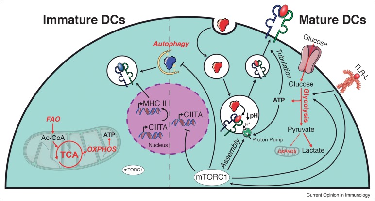
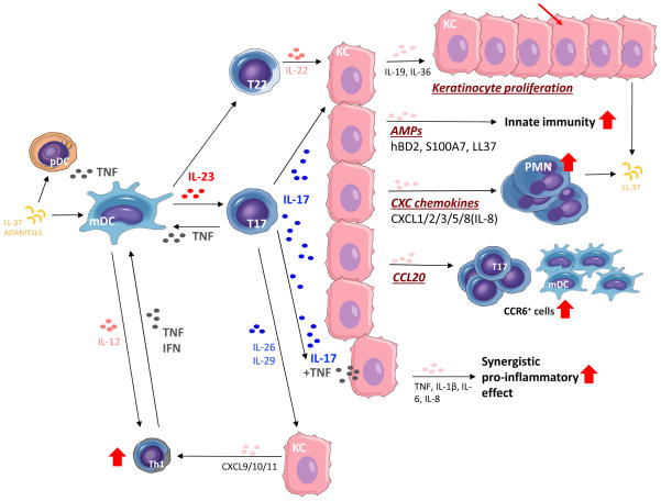
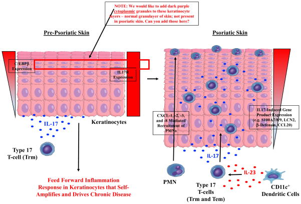


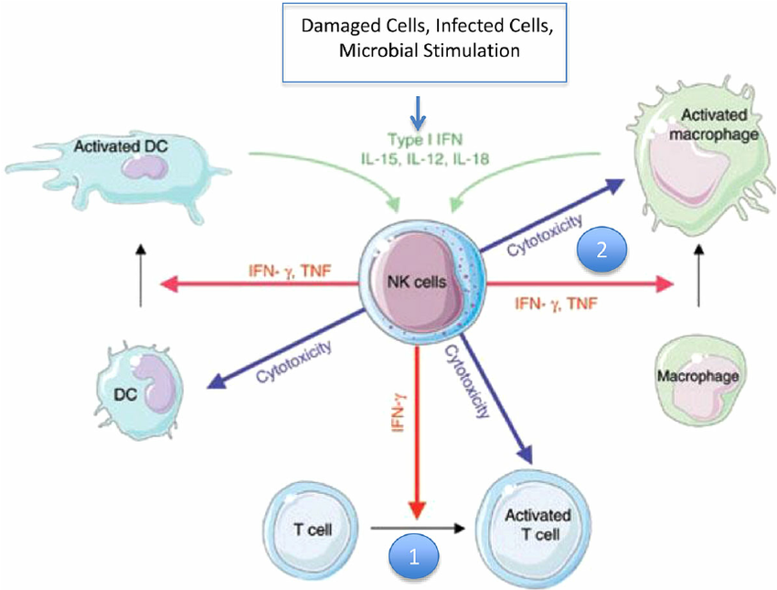
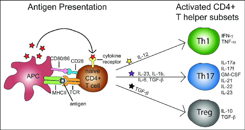
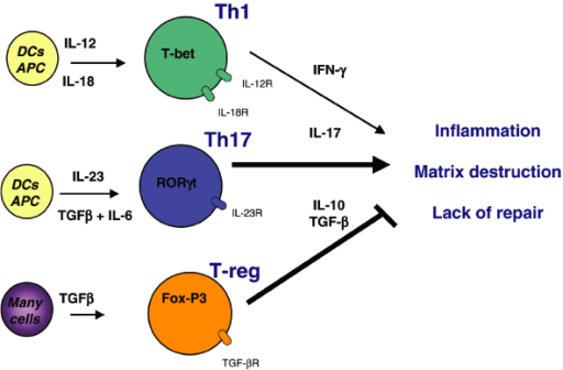
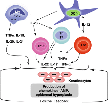
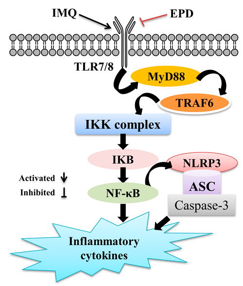
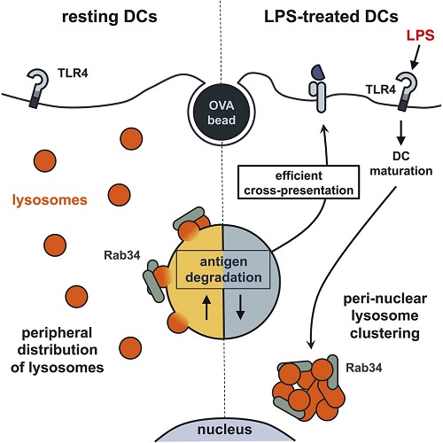
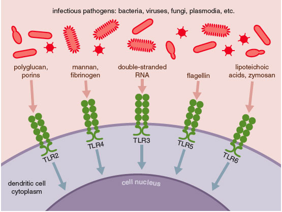
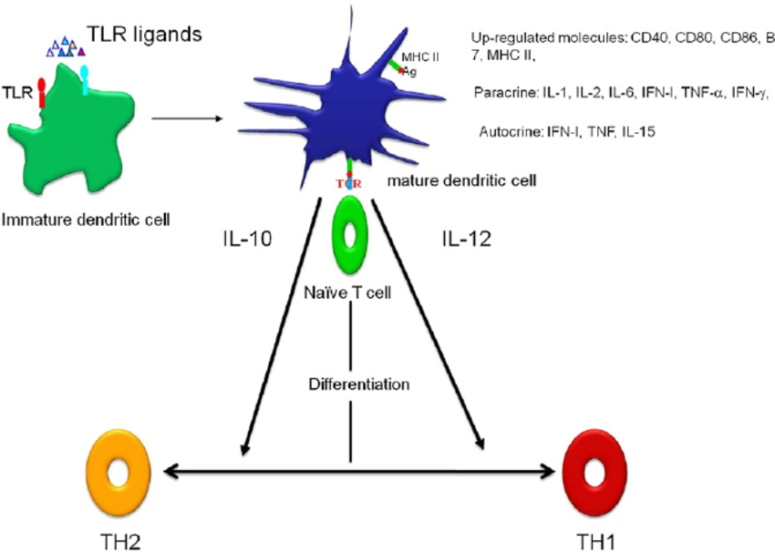
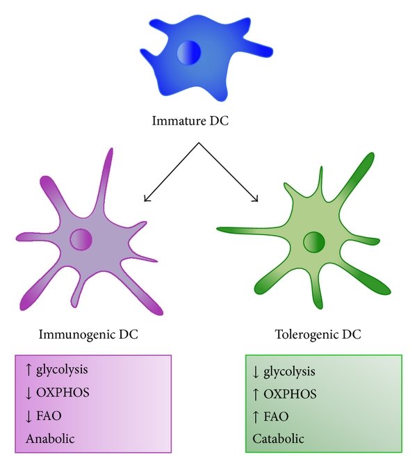
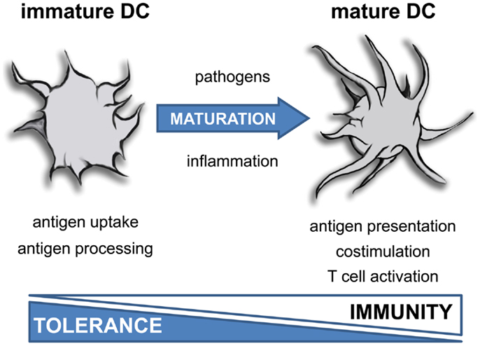
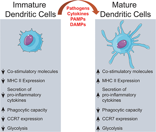
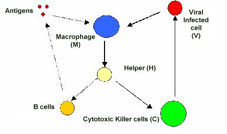

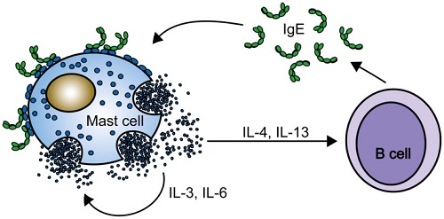
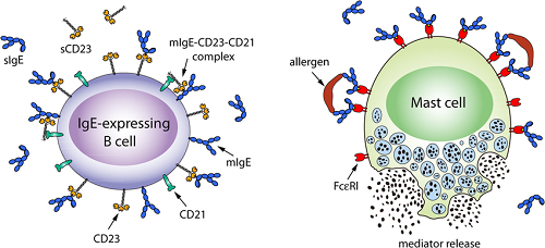


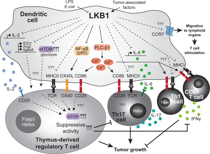
.jpg)