��
��
����ĸ���-Ψһ��������������
liver the only Organ that can Regenerate
��
��
������Ψһ���������������������١���ʵ�ϣ��������ʧ�ߴ�75���ĸ��࣬ʣ�µIJ��ֿ����ٴ������������������࣬����Ĵ����
���������Ǹ����ܹ���ʣ����֯�������ʧ�ĸ�����֯�Ĺ��̡�������Ψһ���������������������١������г����ѧ�˺�������������������֪��ʣ��25����ԭʼ�����������������ص�����ȫ���ߴ硣���鶯�������������Ҫ�Dz�������������Ϊֻ�и�����������滻����������״��Ȼ����������ȵ͵������У�����Ĵ�С����״�����������
����
�������漰��ϸ������Ҫ�Ǹ�ϸ�����ĸ��ƣ�Ȼ��������ϸ�����絨����Ƥϸ�����״��Ƥϸ����һ��ϸ����ֳ��ɣ��·��ѵ�ϸ���������飬Ѫ�����ɺ�ϸ������ʵ����飬������������̡��ڴ��������£��ι��ܽ��ڸ������ڼ��ܵ�����Ӱ�졣��Ȼҩ���л��ijЩ����ܼ��٣������͵�֭���ɵ�����������Ҫ���ܲ�δ�ܵ�ʵ����Ӱ�졣
��������һ���߶��ܿصĹ��̣��ɸ߶������źŵĸ���������ڡ���֪�����ź�ͨ·�̼���������������ϸ�����ӣ��������ӣ����غͺ����塣�����ڷ��ֲ��о���һЩ��Ȼ����ָ��������� - �δ̼����ʣ��������飬����������ǿ����
����
���������������Ǹ�����̬�ĺ��ġ����ڸ�����ҩ��ⶾ����Ҫ��λ���������¶�����ڵ����ѧ���ʣ����ܻ��շ�ϸ�����������ˡ��������Ѹ������������֯���Ӷ���ֹ��������˥�ߡ�Ȼ������������ʵ�ٶȵ�Ԥ������ȡ���ڰ�ϸ������6�Ƿ��й��ȱ�������������ڸ��༲������Ҳ������Ҫ����Ϊ��ά�����������¸��ಿ���г�������ʣ�������������ij����Ʒ���
ʵ��ģ��
������Ҫ���͵�ģ�������о������������������г���Ҳ��Ϊ���ָ��г�����PHX���ͻ�ѧ�յ��ĸ����ˡ�����������ģ���и������Ļ��ƺͶ���ѧ��ͬ����������ͬ���ź�ͨ·�̼�����;���ĸ�������
��
The liver is the only internal organ that can regenerate itself. In fact, you can lose up to 75 percent of your liver, and the remaining parts can regenerate itself into a whole liver again. Amazing stuff.
Liver regeneration is the process by which the liver is able to replace lost liver tissue from growth from the remaining tissue. The liver is the only visceral organ that possesses the capacity to regenerate. The liver can regenerate after either surgical removal or after chemical injury.It is known that as little as 25% of the original liver mass can regenerate back to its full size.The process of regeneration in mammals is mainly compensatory growth because only the mass of the liver is replaced, not the shape.[5] However, in lower species such as fish, both liver size and shape can be replaced.
��
Mechanism
Liver regeneration involves replication of the liver cells, mainly hepatocytes, followed by other cells such as biliary epithelial cells and sinusoidal endothelial cells. Once cell proliferation is completed, the newly divided cells undergo restructuring, angiogenesis and reformation of extracellular matrix to complete the regeneration process.[2] In most cases, liver function is only partially affected during liver regeneration. Whereas certain specialized functions such as drug metabolism decrease, many other primary functions such as albumin and bile production are not substantially affected.[1]
Liver regeneration is a highly controlled process regulated by a complex network on highly redundant signals. Several signaling pathways are known to stimulate regeneration in the liver including cytokines, growth factors, hormones, and nuclear receptors.[1] Discovered and studying in vivo some natural multicomponent liver regeneration substances �C hepatic stimulator substance,[7] hepatic regeneration set,[8] augmenter of liver regeneration.[9]
Function
The ability for the liver to regenerate is central to liver homeostasis. Because the liver is the main site of drug detoxification, it is exposed to many chemicals in the body which may potentially induce cell death and injury. The liver can regenerate damaged tissue rapidly thereby preventing its own failure. However, a predictor of the true speed of liver regeneration depends on whether Interleukin 6 has overexpression[10]. Liver regeneration is also critical for patients of liver diseases where the partial removal of the liver due to fibrosis or tumor is a common therapy that utilizes the ability of the remaining liver to generate back.[citation needed]
Experimental models
Two main types of models are used to study liver regeneration, including surgical removal, also referred to as partial hepatectomy (PHX), and chemical-induced liver damage. Whereas the mechanisms and kinetics of liver regeneration in these two models are different, many of the same signaling pathways stimulate liver regeneration in both pathways.[11]��
��
����������ȱ����Ӱ��-����ĸߴ�л���Ե���~1mL / g / ���ӵĸ߹�ע���ʡ�����Ϣ�����£����Լ��������ѪҺ��Ӧ���ķ�֮һ���ζ����ĸ���ѪҺռ�ܸ����ע���ķ�֮һ�������������������¿��ܻ������ӡ����ӵ�ѪҺ��Ӧʹ���༫���ܵ�����ѭ���ϰ���Ӱ�졣�����˵����س̶Ⱥ�ģʽ��ȡ���ڱ�����Ѫ��ע���ٵ���Թ��ס�
��
��
��
��
��
��
��
��
��
ȱ���Ը�����
ȱ���Ը����˱�����ΪѪ��ת��øˮƽ�Ĵ��������ݵ����ӣ�����������û�����������˼���ԭ�������¸����������֮��IJ�ƽ�⡣
��ͨ�������ڻ����Ҳ��Ѫ������˥�ߺ͵�����������������С��������ذ�������ʧ�����ˮ�ס�֢״���������������̴ٺ����ϸ���ʹ����̫�������ǣ��ڻ������ص���Ѫ֢���Ⱦ���ݿ˵Ļ����пɼ�ȱ���Ը����ˡ������Եأ�ת��øˮƽ����20�������ڼ�����Ѹ�ٻָ�������Ӱ��ѧ�о���ʾ�˵ͻ�������ܶȲ��䣬��Щ����ͨ����ת��ʼ�¼�����ȫ���ˡ����ƺ�Ԥ��ȡ����DZ�ڵļ�����
Liver has high demand on oxygen.
The high metabolic activity of the liver results in a high perfusion rate of ∼1 mL/g/min. Under resting conditions, this is about a quarter of the bodýs total blood supply. The oxygen-rich blood of the hepatic artery contributes to about a quarter of the total liver perfusion that may rise substantially under conditions of excessive oxygen demand. The complex blood supply makes the liver extraordinarily vulnerable to acute circulatory disturbances. Both the severity and the pattern of hepatic injury depend on the relative contribution of passive congestion and diminished perfusion.
Hypoxic Liver Injury
Hypoxic liver injury is defined as a massive, but transient, increase in serum transaminase levels due to an imbalance between hepatic oxygen supply and demand in the absence of other acute causes of liver damage. It typically occurs in elderly individuals with right-sided congestive heart failure and low cardiac output. Precipitating factors include arrhythmias or pulmonary edema. Symptoms include weakness, shortness of breath, and right upper quadrant pain. Less commonly, hypoxic liver injury is seen in patients with severe hypoxemia or septic shock. Characteristically, the transaminase level is elevated 20-fold but normalizes rapidly over several days. Imaging studies reveal hypoechoic or hypodense lesions that resolve completely with reversal of the initiating event. Treatment and prognosis depend on the underlying disease.
��
��
��
��
���ž�����ѪҺ������ѪҺ��ͨ��������Ƣ�������ѪҺ��ͨ�����ž������͵�������нⶾ�� ����������Ըζ����Ķ���Ѫ��Я�������� ��������ѪҺ�Ƕ��صģ���Ϊ����������ѪҺ������ѪҺ����ˣ��ž���ѪҺ��pO2��עѹ����������������١�ѪҺ���ž����ķ�֧ͨ����ΪѪ�ĸ�ϸ�����塱֮��Ŀ�ǻ��ѪҺҲ�Ӹζ����ķ�֧����������Ѫ��������ϸ����Ӧ���������ֻ����ͨ��Ѫ������ռ������뾲���У�������ξ������ξ������������ǻ������
��
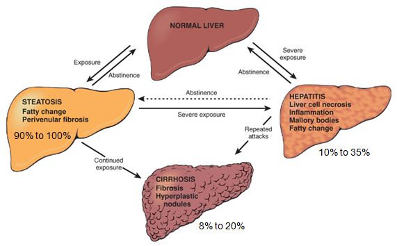
������60~65����ʵ��ϸ��������ϸ����30����35���ķ�ʵ��ϸ����ɣ����ݷ�ϸ����Kupffer cell, KC)������״ϸ����HSC�������Ƥϸ����LSECs����
��
��
��
��
��
��
��
��
REFERENCES
https://en.wikipedia.org/wiki/Liver_regeneration
https://www.ncbi.nlm.nih.gov/pmc/articles/PMC4771376/
https://academic.oup.com/eurheartj/article/34/10/711/496992
https://www.sciencedirect.com/science/article/abs/pii/S0025619611612354
��
��

.png)
.png)
.png)
.png)
.png)
.png)

.png)
.png)
.png)
.png)
.png)
.png)
.png)
.png)
.png)
.png)